Targeting AKT Improves Outcomes in a Subset of Triple Negative Breast Cancer Patients

SUMMARY: Breast cancer is the most common cancer among women in the US and about 1 in 8 women (12%) will develop invasive breast cancer during their lifetime. It is estimated that 252,710 new cases of invasive breast cancer and 63,410 new cases of non-invasive breast cancer will be diagnosed in women in 2017 and 40,610 women are expected to die from the disease. Triple Negative Breast Cancer (TNBC) is a heterogeneous, molecularly diverse group of breast cancers and are ER (Estrogen Receptor), PR (Progesterone Receptor) and HER2 (Human Epidermal Growth Factor Receptor 2) negative. TNBC accounts for 15% to 20% of invasive breast cancers, with a higher incidence noted in young patients. It is usually aggressive, and tumors tend to be high grade and patients with TNBC are at a higher risk of both local and distant recurrence. Those with metastatic disease have one of the worst prognoses of all cancers with a median Overall Survival of 13 months. The majority of patients with TNBC who develop metastatic disease do so within the first 3 years after diagnosis, whereas those without recurrence during this period of time have survival rates similar to those with ER-positive breast cancers. The lack of known recurrent oncogenic drivers in patients with metastatic TNBC, presents a major therapeutic challenge. Nonetheless, patients with TNBC often receive chemotherapy in the neoadjuvant, adjuvant or metastatic settings and approximately 30-40% of patients achieve a pathological Complete Response (pCR) in the neoadjuvant setting. Those who do not achieve a pathological Complete Response tend to have a poor prognosis. It therefore appears that there are subsets of patients with TNBC who may be inherently insensitive to cytotoxic chemotherapy. Three treatment approaches appear to be promising and they include immune therapies, PARP inhibition and inhibition of PI3K pathway.
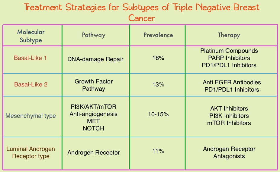
Using gene expression profiling, TNBC can be classified into 4 distinct molecular subtypes- two Basal-Like (BL1, BL2), Mesenchymal type (M) and Luminal Androgen Receptor type (LAR). BL1 molecular subtype of TNBCs are characterized by high levels of expression of genes involved in the cell cycle and DNA-damage repair pathways and accounts for up to 18% of TNBCs. These tumors are more sensitive to therapies targeting the DNA-repair pathways such as platinum based chemotherapy and Poly-ADP Ribose Polymerase (PARP) inhibition. BL2 molecular subtype of TNBCs represent 13% of TNBCs and in contrast are characterized by upregulation of growth factor signaling pathways, including the Epidermal Growth Factor (EGF), MET pathways, as well as genes involved in glycolysis and gluconeogenesis. These tumors may better respond to small molecule inhibitors of growth factor pathways. An alternate classification of Basal-Like subtype includes Basal-Like Immune Suppressed (BLIS) which is associated with downregulation of B cell, T cell, and Natural Killer cell immune-regulating pathways, and has the worse prognosis and Basal-Like Immune Activated (BLIA) subtype, which has the best prognosis due to upregulated immune-associated pathways. Mesenchymal TNBCs constitute approximately 10-30% of TNBC tumors and are associated with aberrations in the PI3K/AKT/ mTOR pathway as well as increased angiogenesis and may benefit from agents targeting these pathways. Metaplastic breast cancer belongs to this TNBC group. The Luminal Androgen Receptor (LAR) subtype accounts for approximately 11% of TNBCs. These tumors have a high expression of Androgen Receptor by IHC (Immuno HistoChemistry) and benefit from Androgen Receptor blockade and do not respond to cytotoxic chemotherapy.

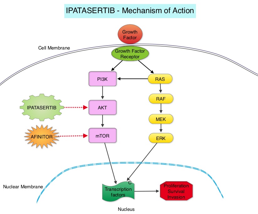
Ipatasertib is a highly selective oral ATP-competitive, small-molecule, AKT inhibitor and sensitivity to Ipatasertib has been associated with high levels of phosphorylated AKT, PTEN protein loss or genetic mutations in PTEN, and PIK3CA mutations. KRAS and BRAF mutations are typically associated with resistance to Ipatasertib. It is estimated that approximately 50% of TNBCs have deficient expression of the tumor suppressor PTEN, which is associated with a higher degree of AKT pathway activation Preclinical studies showed synergy between Ipatasertib and Taxanes. Because of the high prevalence of PI3K/AKT pathway activation in TNBCs, the authors in this study evaluated the benefit of a combination of Ipatasertib and Paclitaxel as first line therapy, for TNBC.
The LOTUS trial is a randomized, placebo controlled, double blind, phase II study in which 124 treatment naïve patients with inoperable, locally advanced or metastatic Triple Negative Breast Cancer were randomly assigned (1:1) to receive Paclitaxel 80 mg/m2 IV Days 1, 8, 15 of a 28 day cycle in combination with either Ipatasertib 400 mg PO daily (N=62) or placebo (N=62), administered on days 1-21 of each 28 day cycle. Treatment was continued until disease progression or unacceptable toxicity. Patients in this study were stratified based on expression of the PTEN tumor suppressor gene and alteration of PIK3CA/AKT1/PTEN in their tumors. The co-primary endpoints were Progression Free Survival (PFS) in the intent-to-treat population and Progression Free Survival in the PTEN-low population. Secondary endpoints included Objective Response Rate and Duration of Response. The median follow up was 10.3 months.

It was noted that the median PFS in the intent-to-treat population was 6.2 months with Ipatasertib versus 4.9 months with placebo (HR=0.60; P=0.037). In the 48 patients with low PTEN expression tumors, the median PFS however was 6.2 months with Ipatasertib and 3.7 months with placebo (HR=0.59; P=0.18), and this was not statistically significant. The PFS benefit was more pronounced in the patient group with PIK3CA/AKT1/PTEN-altered tumors, with a median PFS of 9.0 months in the Ipatasertib group versus 4.9 months in the placebo group (HR=0.44; P=0.041). The most common grade 3/4 adverse events in the Ipatasertib group were diarrhea and neutropenia.

The authors concluded that Ipatasertib prolonged Progression Free Survival compared to placebo and is the first study supporting AKT-targeted therapy for Triple Negative Breast Cancer, supporting the use of gene expression profiling in this heterogeneous malignancy. Ipatasertib plus paclitaxel versus placebo plus paclitaxel as first-line therapy for metastatic triple-negative breast cancer (LOTUS): a multicentre, randomised, double-blind, placebo-controlled, phase 2 trial. Kim S, Dent R, Im S, et al on behalf of the LOTUS investigators. The Lancet Oncology 2017;18:1360-1372
FDA Approves OPDIVO® for Advanced Hepatocellular Carcinoma

SUMMARY: The FDA on September 22, 2017, granted accelerated approval to OPDIVO® (Nivolumab) for the treatment of HepatoCellular Carcinoma (HCC), in patients who have been previously treated with NEXAVAR® (Sorafenib). The American Cancer Society estimates that about 41,000 new cases of primary liver cancer will be diagnosed in the US for 2017 and 29,000 patients will die of their disease. Liver cancer is seen more often in men than in women and the incidence has more than tripled since1980. This increase has been attributed to the higher rate of hepatitis C virus (HCV) infection among baby boomers (born between 1945 through 1965). Obesity and type II diabetes have also likely contributed to the trend. Other risk factors include alcohol, which increases liver cancer risk by about 10% per drink per day, and tobacco use, which increases liver cancer risk by approximately 50%. HCC is the second most common cause of cancer-related deaths worldwide. NEXAVAR® (Sorafenib) was approved by the FDA in 2007 for the treatment of unresectable, HepatoCellular Carcinoma (HCC). Patients with advanced HCC who progress on NEXAVAR®, have a poor prognosis with limited treatment options.

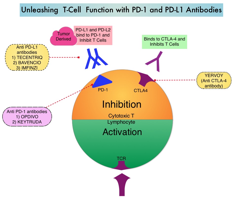
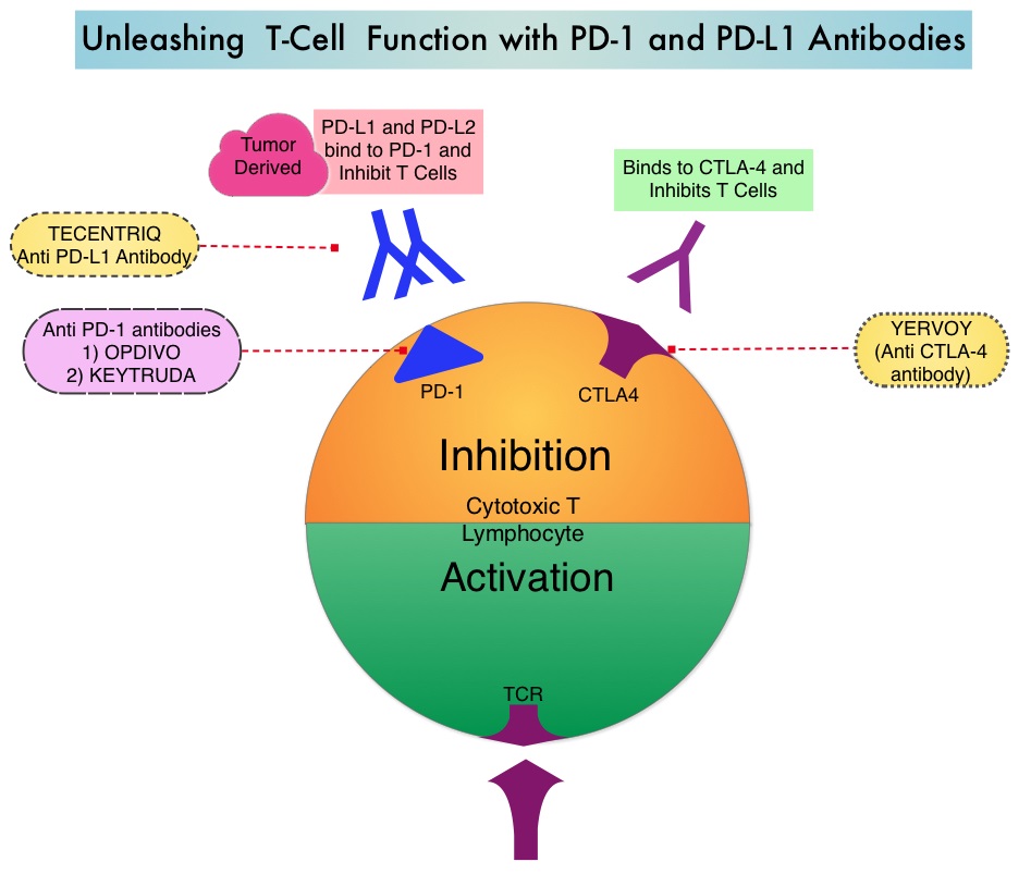
Immune checkpoints are cell surface inhibitory proteins/receptors that are expressed on activated T cells. They harness the immune system and prevent uncontrolled immune reactions. Under normal circumstances, Immune checkpoints or gate keepers inhibit intense immune responses by switching off the T cells of the immune system and can thereby suppress antitumor immunity. These Immune checkpoint proteins/receptors include CTLA-4 (Cytotoxic T-Lymphocyte Antigen 4, also known as CD152), PD-1(Programmed cell Death 1), etc. Immune check point inhibitors are antibodies that target the immune checkpoint receptors and unleash the T cells, resulting in T cell proliferation, activation and a therapeutic response. OPDIVO® (Nivolumab) is an immune checkpoint PD-1 (Programmed cell Death 1) targeted, fully human, immunoglobulin G4 monoclonal antibody that demonstrated significant activity in HepatoCellular Carcinoma (HCC).

The approval of OPDIVO® in HCC was based on a 154-patient subgroup of CHECKMATE-040 study, which is a multicenter, open-label trial, conducted in patients with HCC and Child-Pugh A cirrhosis, who had progressed on or were intolerant to NEXAVAR®. Enrolled patients included patients without active viral hepatitis, patients with either active HBV-Hepatitis B Virus (31%) or HCV- Hepatitis C Virus (21%). Patients with active co-infection with HBV and HCV or with Hepatitis D Virus infection were excluded. Patients received OPDIVO® 3 mg/kg by IV infusion every 2 weeks. The median age was 64 years and 80% of patients were male. The primary endpoints were safety/tolerability and Objective Response Rate (ORR) by investigator and Blinded Independent Central Review. Secondary endpoints included Duration of Response, Disease Control Rate and Overall Survival.

The confirmed Objective Response Rate as assessed by Independent Central Review was 19 % with 3 Complete Responses and 19 partial responses(13%) and 41% of the patients had stable disease. Response duration ranged from 3 months to over 38 months and 91% of responders had responses lasting 6 months or longer and 55% of the patients had responses lasting 12 months or longer. Overall responses were noted regardless of tumor cell PD-L1 expression and chronic hepatitis virus infection. Common adverse reactions included fever, rash, musculoskeletal pain, nausea, diarrhea, cough and dyspnea. There was a higher incidence of elevations in transaminases and bilirubin levels noted.

It was concluded that OPDIVO® demonstrated durable responses with long-term survival and favorable safety in patients with advanced HCC. This FDA approval provides a new treatment option for appropriate patients with HCC, who progress on prior systemic therapy. STIVARGA® (Regorafenib) is another agent presently available for this subset of patients. Nivolumab (nivo) in sorafenib (sor)-naive and -experienced pts with advanced hepatocellular carcinoma (HCC): CheckMate 040 study. Crocenzi TS, El-Khoueiry AB, Yau TC, et al. J Clin Oncol. 2017;35 (suppl; abstr 4013).

African American Women with Benign Breast Disease and Triple Negative Breast Cancer

SUMMARY: Breast cancer is the most common cancer among women in the US and about 1 in 8 women (12%) will develop invasive breast cancer during their lifetime. It is estimated that 252,710 new cases of invasive breast cancer and 63,410 new cases of non-invasive breast cancer will be diagnosed in women in 2017 and 40,610 women are expected to die from the disease. Benign Breast Disease (BBD) is an important risk factor for the later development of breast cancer in either breast. With the routine use of mammography, the identification of BBD has become more common, and this has in turn enabled the calculation of breast cancer risk. The Breast Cancer Risk Assessment Tool which is an interactive tool designed by researchers at the National Cancer Institute (NCI) and the National Surgical Adjuvant Breast and Bowel Project (NSABP) to estimate a woman's risk of developing invasive breast cancer, utilizes the number of breast biopsies and atypical hyperplasia, in addition to other factors, to estimate women’s risk of developing breast cancer. Benign Breast Disease (BBD) encompasses a variety of histologic entities. FibroCystic Changes (FCCs) constitute the most frequent benign disorder of the breast. FCCs may be multifocal and bilateral and women usually present with symptoms of breast pain and tender nodularities in breasts. Hormonal imbalance, with estrogen predominance over progesterone, has been implicated as an important contributing factor in its development. Fibrocystic changes are usually subdivided into nonproliferative lesions, proliferative lesions without atypia, and proliferative lesions with atypia (atypical hyperplasia). Proliferative or atypical lesions are associated with an increased risk of breast cancer. However a significant majority of breast biopsies (about 70%) show nonproliferative lesions.

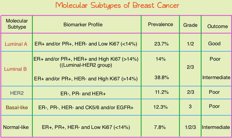
Breast cancer is heterogeneous malignancy and using global gene expression analyses, 6 breast cancer intrinsic subtypes have been established. They include Luminal A, Luminal B, HER2-enriched, Claudin-low, Basal-like, and a Normal breast-like group. Triple Negative Breast Cancer (TNBC) accounts for 12–24% of all breast carcinomas and is negative for Estrogen Receptor (ER), Progesterone Receptor (PR), and HER2. There is a 2 fold increase in the incidence of TNBC among African American (AA) women compared with White American (WA) women and is a surrogate for the inherently aggressive Basal-like breast cancer subtype. This group has the worse prognosis compared to other breast cancer subtypes. Basal-like breast cancer sub-type is also a marker of hereditary breast cancer susceptibility. Multiparity may increase the risk of TNBC and reduce the likelihood of developing ER-positive breast cancer. Triple Negative Breast Cancer (TNBC) compared with non-TNBC, likely arises from different pathogenetic pathways. NF-κB pathway, which controls immune response, angiogenesis, the cell cycle, extracellular matrix degradation, and apoptosis, may represent a key regulator of TNBC. Activation of PI3K/AKT pathway in turn, has been implicated in oncogenic transformation.

Benign breast disease (BBD) has been associated with increased risk for ER positive/non-TNBC. The purpose of this study was to determine whether African American (AA) identity is associated with TNBC among a cohort of both AA and White American (WA) women, who were initially diagnosed with BBD. The authors in this study conducted a retrospective analysis of a cohort comprising 2588 African American women and 3566 White American women aged between 40 and 70 years, with a biopsy-proven Benign Breast Disease. This data was obtained from the Pathology Information System of Henry Ford Health System (HFHS), an integrated multihospital and multispecialty health care system in Detroit, Michigan. These individuals had breast biopsies performed between January 1, 1994, and December 31, 2005 and data analysis was performed from November 1, 2015, to June 15, 2016. Patients with prior breast cancer and those with breast cancer diagnosed within 6 months of BBD biopsy were excluded. Benign Breast Disease was classified as fibrocystic/proliferative/hyperplasia without atypia, with atypia, or with lobular carcinoma in situ. The mean follow up was 10.2 years for both AA patients and WA patients.

It was noted that more than 75% of the subsequent breast cancers in each subset were Ductal Carcinoma in Situ (DCIS) or Stage I. Among the African American (AA) patients who developed subsequent invasive breast cancer, 24.2% developed TNBC compared with 7.4% of the White American (WA) patients who developed subsequent invasive breast cancers (P=0.01). The 10 year probability estimate for developing TNBC was 0.56% for AA patients and 0.25% for WA patients.

The authors concluded that in this largest analysis to date of TNBC and its relationship to racial/ethnic identity and Benign Breast Disease as risk factors, African American identity persisted as a significant risk factor for Triple Negative Breast Cancer, suggesting that African American identity is associated with inherent susceptibility for TNBC pathogenetic pathways. This important finding should be taken into consideration, when discussing chemoprevention strategy among African American women with Benign Breast Diseases. Association Between Benign Breast Disease in African American and White American Women and Subsequent Triple-Negative Breast Cancer. Newman LA, Stark A, Chitale D, et al. JAMA Oncol. 2017;3:1102-1106.

FDA Approves TAFINLAR® and MEKINIST® Combo for BRAF V600E-Mutant Non Small Cell Lung Cancer

SUMMARY: The FDA on June 22, 2017 granted regular approvals to TAFINLAR® ((Dabrafenib) and MEKINIST® (Trametinib) administered in combination, for patients with metastatic Non Small Cell Lung Cancer (NSCLC) with BRAF V600E mutation, as detected by an FDA-approved test. These are the first FDA approvals specifically for treatment of patients with BRAF V600E mutation-positive metastatic NSCLC. The FDA also approved the Oncomine® Dx Target Test (Thermo Fisher Scientific), a Next Generation Sequencing (NGS) test to detect multiple gene mutations for lung cancer in a single test from a single tissue specimen. This test detects the presence of BRAF, ROS1, and EGFR gene mutations or alterations in tumor tissue of patients with NSCLC. This test can be used to select patients with NSCLC with the BRAF V600E mutation for treatment with the combination of TAFINLAR® and MEKINIST®. This is the first NGS oncology panel test approved by the FDA for multiple companion diagnostic indications. Lung cancer is the second most common cancer in both men and women and accounts for about 13% of all new cancers and 27% of all cancer deaths. The American Cancer Society estimates that for 2017 about 222,500 new cases of lung cancer will be diagnosed and over 155,000 patients will die of the disease. Lung cancer is the leading cause of cancer-related mortality in the United States.

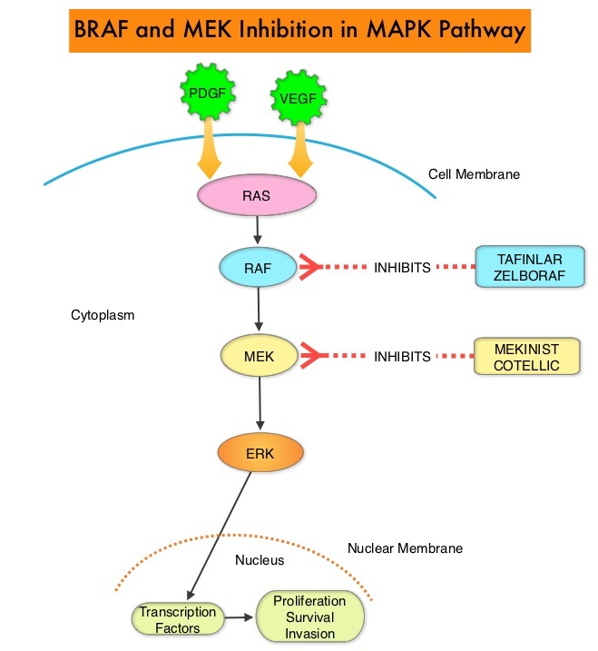
The approval of the combination of MEKINIST® (Trametinib) and TAFINLAR® (Dabrafenib), to treat patients with advanced NSCLC, was based on the understanding of the biological pathways of this malignancy. The Mitogen-Activated Protein Kinase pathway (MAPK pathway) is an important signaling pathway which enables the cell to respond to external stimuli. This pathway plays a dual role, regulating cytokine production and participating in cytokine dependent signaling cascade. The MAPK pathway of interest is the RAS-RAF-MEK-ERK pathway. The RAF family of kinases includes ARAF, BRAF and CRAF signaling molecules. BRAF is a very important intermediary of the RAS-RAF-MEK-ERK pathway. BRAF mutations have been demonstrated in 6%-8% of all malignancies and BRAF V600E mutation occurs in 1-2% of lung adenocarcinomas and acts as an oncogenic driver.

BRF113928 is an open-label, multicohort, multicentre, non-randomized, phase II study which sequentially enrolled patients with BRAF V600E mutation-positive metastatic NSCLC, across 3 cohorts. The first 2 cohorts included previously treated patients. The median age was 64 years, 98% had adenocarcinoma, and majority of patients (72%) were former or current smokers. In the first cohort, 84 patients received single agent TAFINLAR® following one or more prior platinum-based chemotherapy. In the second cohort, 57 patients received a combination of TAFINLAR® 150 mg orally twice daily and MEKINIST® 2 mg orally once daily. Patients in this second cohort had received at least 1 prior line of platinum-based chemotherapy regimen with disease progression and 33% had received 2 or more prior chemotherapy regimens.

It was noted that in these previously treated cohorts of patients, the Objective Response Rate (ORR) for the combination treatment based on independent review was 63%, with a median Duration of Response (DoR) of 12.6 months. The ORR for patients who received single agent TAFINLAR® was 27% and the median DoR was 9.9 months. The investigator assessed median Progression Free Survival (PFS) was 10.2 months, and median Overall Survival (OS) was 18.2 months.

The authors in this latest publication reported the the efficacy and safety of TAFINLAR® plus MEKINIST® treatment in previously untreated patients with BRAF V600E-mutant metastatic NSCLC (third cohort). This cohort enrolled 36 patients and patients received TAFINLAR® 150 mg orally twice daily plus MEKINIST® 2 mg orally once daily, until disease progression or unacceptable toxicities. The median follow up was 15.9 months. The Primary endpoint was investigator assessed ORR. Secondary endpoints included Duration of Response (DoR), PFS, OS, and safety.

The Objective Response Rate in this cohort was 64%, with 6% Complete Response and 58% Partial Response. The Disease Control Rate was 75%. The median Duration of Response was 10.4 months, median PFS was 10.9 months and median OS was 24.6 months. The most common grade 3 or 4 adverse events were pyrexia, liver function abnormalities, hypertension and vomiting.

The authors concluded that TAFINLAR® plus MEKINIST® demonstrated substantial antitumor activity and durable responses in patients with treatment-naive BRAF V600E-mutant NSCLC. This study confirmed that there is a fourth actionable biomarker, BRAF V600E, in addition to EGFR, ALK and ROS-1, for patients with Non Small Cell Lung Cancer. Dabrafenib plus trametinib in patients with previously untreated BRAF V600E-mutant metastatic non-small-cell lung cancer: an open-label, phase 2 trial. Planchard D, Smit EF, Groen HJ, et al. The Lancet Oncology 2017;18:1307-1316

FDA Approves VERZENIO® for Hormone Receptor Positive, HER2-Negative Breast Cancer

SUMMARY: The FDA on September 28, 2017, approved VERZENIO® (Abemaciclib) in combination with FASLODEX® (Fulvestrant) for women with Hormone Receptor positive (HR-positive), HER2-negative, advanced or metastatic breast cancer, with disease progression following endocrine therapy. In addition, VERZENIO® was approved as monotherapy for women and men with HR-positive, HER2-negative advanced or metastatic breast cancer with disease progression following endocrine therapy and prior chemotherapy in the metastatic setting. Breast cancer is the most common cancer among women in the US and about 1 in 8 women (12%) will develop invasive breast cancer during their lifetime. It is estimated that 252,710 new cases of invasive breast cancer and 63,410 new cases of non-invasive breast cancer will be diagnosed in women in 2017 and 40,610 women are expected to die from the disease.

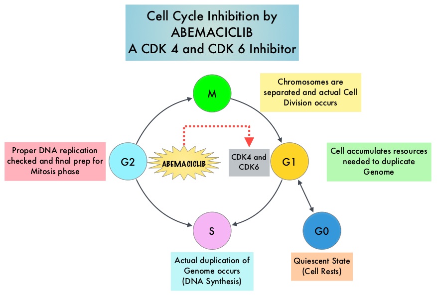
Cyclin Dependent Kinases (CDK) play a very important role to facilitate orderly and controlled progression of the cell cycle. Genetic alterations in these kinases and their regulatory proteins have been implicated in various malignancies. Cyclin Dependent Kinases 4 and 6 (CDK4 and CDK6), phosphorylate RetinoBlastoma protein (RB), and initiate transition from the G1 phase to the S phase of the cell cycle. RetinoBlastoma protein has antiproliferative and tumor-suppressor activity and phosphorylation of RB protein nullifies its beneficial activities. CDK4 and CDK6 are activated in hormone receptor positive breast cancer, promoting breast cancer cell proliferation. Further, there is evidence to suggest that endocrine resistant breast cancer cell lines depend on CDK4 for cell proliferation. The understanding of the role of Cyclin Dependent Kinases in the cell cycle, has paved the way for the development of CDK inhibitors.

VERZENIO® is an oral, selective inhibitor of CDK4 and CDK6 kinase activity, and prevents the phosphorylation and subsequent inactivation of the Rb tumor suppressor protein, thereby inducing G1 cell cycle arrest and inhibition of cell proliferation. VERZENIO® is structurally distinct from other CDK 4 and 6 inhibitors (such as Ribociclib and Palbociclib) and is 14 times more potent against cyclin D1/CDK 4 and cyclin D3/CDK 6, in enzymatic assays. VERZENIO® in the phase I trials was noted to be active in HR-positive, metastatic breast cancer, as monotherapy as well as in combination with FASLODEX®. Based on this preliminary data, two clinical trials, MONARCH 1 and MONARCH 2, were conducted.

The approval of VERZENIO® as monotherapy in HR-positive, metastatic breast cancer was based on MONARCH 1, which is a a single-arm, open-label, phase II, multicenter study, which enrolled women with measurable HR-positive, HER2-negative metastatic breast cancer, whose disease progressed during or after endocrine therapy, had received a taxane in any setting, and who received one or two prior chemotherapy regimens in the metastatic setting. This trial included 132 patients who received VERZENIO® 200 mg orally twice daily on a continuous schedule, until disease progression. The Objective Response Rate was 19.7%, with a median response duration of 8.6 months.

The approval of VERZENIO® in combination with FASLODEX® was based on MONARCH 2, which is an international, double-blind, phase III study in which 669 patients were randomized in a 2:1 ratio to receive either VERZENIO® plus FASLODEX® (N=446) or placebo plus FASLODEX® (N=223). Enrolled patients had HR-positive, HER2-negative metastatic breast cancer, with disease progression while receiving neoadjuvant or adjuvant endocrine therapy, within 12 months of adjuvant endocrine therapy, or while receiving endocrine therapy for metastatic breast cancer. Patients must not have received more than one endocrine therapy or any prior chemotherapy for metastatic breast cancer. Randomized patients received either VERZENIO® 150 mg or placebo orally twice daily plus FASLODEX® 500 mg IM on Day 1 and Day 15 of cycle 1 and then on Day 1 of cycle 2 and beyond (28-day cycles). Treatment was continued until disease progression or unmanageable toxicities. The mean patient age was 60 years, 82% of patients were postmenopausal, 72% had measurable disease, 56% had visceral disease, and 25% had primary endocrine therapy resistance. About 60% of patients had received chemotherapy in the adjuvant or neoadjuvant setting and 69% of the patients had prior therapy with Aromatase Inhibitors (AI). The Primary end point was Progression Free Survival (PFS), and Secondary end points included Overall Survival (OS), Objective Response Rate (ORR), Duration of Response, Clinical Benefit Rate, Quality of Life, and safety.

The median PFS for the group receiving VERZENIO® plus FASLODEX® was 16.4 months compared with 9.3 months for those taking placebo with FASLODEX® (HR= 0.55; P<0.0001). In patients with measurable disease, the Objective Response Rate for the group receiving VERZENIO® plus FASLODEX® was 48.1% compared to 21.3% in the placebo with FASLODEX® treated patients. The most common adverse events in the VERZENIO® versus placebo groups were diarrhea neutropenia, nausea and fatigue.

It was concluded that a combination of VERZENIO® plus FASLODEX® significantly improved Progression Free Survival and Objective Response Rates, with a tolerable safety profile, in patients with Hormone Receptor-positive and HER 2-negative metastatic breast cancer who progressed while receiving endocrine therapy. MONARCH 2: Abemaciclib in Combination With Fulvestrant in Women With HR+/HER2− Advanced Breast Cancer Who Had Progressed While Receiving Endocrine Therapy. Sledge GW, Toi M, Neven P, et al. DOI: 10.1200/JCO.2017.73.7585 Journal of Clinical Oncology 35, no. 25 (September 2017) 2875-2884.

MONARCH 1: Results from a phase II study of abemaciclib, a CDK4 and CDK6 inhibitor, as monotherapy, in patients with HR+/HER2- breast cancer, after chemotherapy for advanced disease. Dickler MN, Tolaney SM, Rugo HS, et al. J Clin Oncol 34, 2016 (suppl; abstr 510).

FDA Approves ALIQOPA®, a PI3K Inhibitor, for Follicular Lymphoma

SUMMARY: The FDA on September 14, 2017, granted accelerated approval to ALIQOPA® (Copanlisib) for the treatment of adult patients with relapsed Follicular Lymphoma, who have received at least two prior systemic therapies. The American Cancer Society estimates that in 2017, about 72,240 people will be diagnosed with Non Hodgkin Lymphoma (NHL) in the United States and about 20,140 individuals will die of this disease. Indolent NHLs are mature B cell lymphoproliferative disorders and include Follicular Lymphoma, Nodal Marginal Zone Lymphoma (NMZL), Extranodal Marginal Zone Lymphoma (ENMZL) of Mucosa-Associated Lymphoid Tissue (MALT), Splenic Marginal Zone Lymphoma (SMZL), LymphoPlasmacytic Lymphoma (LPL) and Small Lymphocytic Lymphoma (SLL). Follicular Lymphoma is the most indolent form and second most common form of all NHLs and they are a heterogeneous group of lymphoproliferative malignancies. Approximately 20% of all NHLs are Follicular Lymphomas. Advanced stage indolent NHL is not curable and as such, prolonging Progression Free Survival (PFS) and Overall Survival (OS), while maintaining Quality of Life, have been the goals of treatment intervention. Asymptomatic patients with indolent NHL are generally considered candidates for “watch and wait” approach whereas those with B symptoms (fever, night sweats, and weight loss), painful lymphadenopathy/splenomegaly, organ compromise and cytopenias are generally considered candidates for therapy.. Follicular Lymphoma International Prognostic Index (FLIPI) is of prognostic value and is used to help with treatment choices. The Ann Arbor classification divides FL into four stages. Patients with stages I and II have localized disease and those with stages III and IV have advanced disease. The World Health Organization (WHO) further classified FL based on histology into low grade (grades 1 and 2) and high grade (grade 3a) FLs. Grade 3b FL which demonstrates diffuse areas of involvement is designated as Diffuse Large B-cell Lymphoma (DLBCL) and is treated as such. Patients with advanced stage symptomatic Follicular Lymphoma are often treated with induction chemoimmunotherapy followed by maintenance RITUXAN® (Rituximab). This can result in a median PFS of 6-8 yrs and a median Overall Survival of 12-15 yrs. However, approximately 30% of the patients will relapse in 3 years and treatment options are limited for patients with relapses, after multiple treatments.

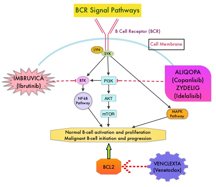
ALIQOPA® is a pan-class 1, PI3K inhibitor with inhibitory activity predominantly against PI3K-α and PI3K-δ Isoforms expressed in malignant B cells. The alpha isoform is broadly expressed and involved in insulin signaling and angiogenesis, as well as resistance mechanisms to lymphoma whereas the delta isoform is expressed by leukocytes and is involved in B-cell signaling, development, and survival. ALIQOPA® has been shown to induce tumor cell death by apoptosis and inhibition of proliferation of primary malignant B cell lines. ALIQOPA® also inhibits several key cell-signaling pathways, including B-cell receptor (BCR) signaling, CXCR12 (C-X-C chemokine receptor 12) mediated chemotaxis of malignant B cells, and NFϰB (Nuclear Factor Kappa B) signaling in lymphoma cell lines.

The approval of ALIQOPA® was based on data from the CHRONOS-1 trial, which is an open-label, single arm, multicenter, phase II study of patients with relapsed, Indolent or aggressive Non Hodgkin Lymphomas. This trial included patients with Follicular lymphoma (Grades 1-3a), Marginal Zone Lymphoma, Small Lymphocytic Lymphoma, and LymphoPlasmacytic Lymphoma /Waldenstrom Macroglobulinemia . Eligible patients had relapsed or refractory disease and had received at least two prior systemic therapies. The efficacy data leading to the FDA approval included 104 patients with Follicular B-cell Non Hodgkin Lymphoma. In this trial, patients received 0.8 mg/kg or 60 mg of ALIQOPA® by IV infusion on days 1, 8, and 15 of a 28-day treatment cycle, until disease progression or development of unacceptable toxicity. The median patient age was 63 yrs and all study patients had prior exposure to RITUXAN® and one or more alkylating agents, and 60% of the patients had disease that was refractory to the last regimen received. The Primary endpoint was Objective Response Rate after a minimum of 16 weeks of treatment. Secondary endpoints included Progression Free Survival, Duration of Response, Overall Survival, safety, and Quality of Life.

The Objective Response Rate was 58.7%, with an estimated median response duration of 12.2 months. The Complete Response rate was 14.4% and partial response rate was 44.3% and 33.7% has stable disease. The most common adverse reactions included nausea, hyperglycemia, diarrhea, fatigue, hypertension, cytopenias and lower respiratory tract infections. According to the authors, safety was manageable compared with the other PI3K inhibitor approved by the FDA, ZYDELIG® (Idelalisib), which targets only the δ-isoform and has warnings against fatal or severe colitis, intestinal perforation, hepatotoxicity, and pneumonitis The safety advantage with ALIQOPA® may be due to the dose scheduling or the IV mode of delivery.

It was concluded that ALIQOPA® has significant activity in patients with relapsed/refractory indolent B-cell lymphoma, and the safety was manageable, compared with other PI3K inhibitors. Two phase III trials are underway, using ALIQOPA® in combination with RITUXAN®. Dreyling M, Santoro A, Mollica L, et al. Copanlisib in patients with relapsed or refractory indolent B-cell lymphoma: Primary results of the pivotal CHRONOS-1 study. Presented at: 2017 AACR Annual Meeting; April 1-5, 2017; Washington, DC. Abstract CT149.

FDA Approves KEYTRUDA® for Advanced Gastric Cancer

SUMMARY: The FDA on September 22, 2017 granted accelerated approval to KEYTRUDA® (Pembrolizumab), for patients with recurrent locally advanced or metastatic, Gastric or GastroEsophageal junction adenocarcinoma, whose tumors express PD-L1, as determined by an FDA-approved test. Patients must have had disease progression on or after two or more prior systemic therapies, including Fluoropyrimidine and platinum-containing chemotherapy and, if appropriate, HER2/neu-targeted therapy. Cancers of the esophagus and stomach are among the most prevalent malignancies and are a major cause of cancer-related mortality. It is estimated that in 2016 GastroEsophageal Adenocarcinoma (GEA) accounted for 43,280 new cases in the United States. The incidence of these tumors has been on the rise in the past decade. Majority of the patients with GEA have advanced disease at the time of initial presentation and have limited therapeutic options with little or no chance for cure. Patients with localized disease (stage II and stage III) are often treated with multimodality therapy and 40% of the patients may survive for 5 years or more.

KEYTRUDA® is a fully humanized, Immunoglobulin G4, anti-PD-1, monoclonal antibody, that binds to the PD-1 receptor and blocks its interaction with ligands PD-L1 and PD-L2, thereby undoing PD-1 pathway-mediated inhibition of the immune response and unleashing the tumor-specific effector T cells. In previously reported Phase I study, KEYTRUDA® showed promising antitumor activity with manageable safety, in patients with previously treated advanced gastric cancer.

Based on these findings, the authors conducted a global, multicohort, Phase II study (KEYNOTE 059), in which patients with advanced gastric or gastroesophageal junction cancer received KEYTRUDA® 200 mg every 3 weeks for up to 2 years or until disease progression or unacceptable toxicity. Cohort 1 in this study enrolled 259 patients of whom 57% (N=148) had PD-L1 positive tumors and these tumors were either MicroSatellite Stable (MSS), or had undetermined MicroSatellite Instability (MSI) or MisMatch Repair (MMR) status. PD-L1 expression was evaluated by the PD-L1 IHC (22C3 antibody) and PD-L1 positivity was based on a Combined Positive Score (CPS) of 1 or more. CPS is determined by the number of PD-L1 staining cells (tumor cells, lymphocytes, macrophages) divided by total number of tumor cells evaluated, multiplied by 100. The median age was 62 years, 76% were men and over 50% of the patients received KEYTRUDA® as third line treatment or beyond. The median duration of follow up was 5.4 months. Primary end points included Objective Response Rate (ORR), safety, and tolerability.

In the group of patients with PD-L1 positive tumors, the ORR was 15.5% with 2% Complete Responses and 13.5% Partial Responses. Among the responding patients, the response duration ranged from 3-19 months, with 58% of the responding patients having response durations of 6 months or longer and 26% of the responding patients having response durations of 12 months or longer. In this cohort of 259 patients, 3% had tumors that were determined to be MSI-High. The ORR in this group was 57%, with response duration ranging from 5-14 months. In the PD-L1 negative patients however, the ORR was only 5.5%.

It was concluded that KEYTRUDA® demonstrated promising antitumor activity and durable responses in patients with advanced Gastric/GastroEsophageal Junction cancer, who had progressed on more than 2 lines of therapy, with higher Objective Response Rates noted in patients with PD-L1-positive tumors. KEYNOTE-059 cohort 1: Efficacy and safety of pembrolizumab (pembro) monotherapy in patients with previously treated advanced gastric cancer. Fuchs CS, Doi T, Jang RW-J, et al. J Clin Oncol. 2017;35 (suppl; abstr 4003).

FDA Approves First Biosimilar for Cancer Treatment

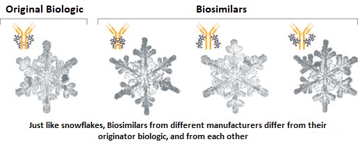
SUMMARY: The FDA on Sept. 14, 2017 approved MVASI® (Bevacizumab-awwb) as a Biosimilar to AVASTIN® (Bevacizumab). Bevacizumab is a recombinant immunoglobulin G1 (IgG1) monoclonal antibody (mAb) that binds to Vascular Endothelial Growth Factor (VEGF) and inhibits the interaction of VEGF with its receptors, VEGF receptor-1 and VEGF receptor-2. This in turn inhibits establishment of new blood vessels that are essential for the maintenance and growth of solid tumors. MVASI® is the first Biosimilar approved in the U.S. for the treatment of cancer. A Biosimilar product is a biological product that is approved based on its high similarity to an already approved biological product (also known as reference product). Biological products are made from living organisms including humans, animals and microorganisms such as bacteria or yeast, and are manufactured through biotechnology, derived from natural sources or produced synthetically. Biological products have larger molecules with a complex structure, than conventional drugs (also known as small molecule drugs).

Unlike biological products, conventional drugs are made of pure chemical substances and their structures can be identified. A generic drug is a copy of brand name drug and has the same active ingredient and is the same as brand name drug in dosage form, safety and strength, route of administration, quality, performance characteristics and intended use. Therefore, brand name and the generic drugs are bioequivalent. The Affordable Care Act in 2010 created an abbreviated licensure pathway for biological products that are demonstrated to be “Biosimilar” to, or “interchangeable” with an FDA approved biological product (reference product). The Biosimilar must show that it has no clinically meaningful differences in terms of safety and effectiveness from the reference product. A Biosimilar product can only be approved by the FDA if it has the same mechanism of action, route of administration, dosage form and strength as the reference product, and only for the indications and conditions of use that have been approved for the reference product. Biosimilars are not as easy to manufacture as generics (copies of brand name drugs), because of the complexity of the structure of the biologic product and the process used to make a biologic product. The facilities where Biosimilars are manufactured must also meet the FDA standards.

MVASI® is approved for the treatment of patients with the following cancers:

• Metastatic Colorectal cancer, in combination with intravenous 5-Fluorouracil-based chemotherapy for first or second line treatment. MVASI® is not indicated for the adjuvant treatment of surgically resected Colorectal cancer.

• Metastatic Colorectal cancer, in combination with Fluoropyrimidine-Irinotecan or Fluoropyrmidine-Oxaliplatin-based chemotherapy for the second line treatment of patients who have progressed on a first-line Bevacizumab containing regimen.

• Non-squamous Non Small Cell Lung Cancer, in combination with Carboplatin and Paclitaxel for first line treatment of unresectable, locally advanced, recurrent or metastatic disease.

• Glioblastoma with progressive disease following prior therapy, based on improvement in Objective Response Rate. No data is available demonstrating improvement in disease-related symptoms or survival with Bevacizumab.

• Metastatic Renal cell carcinoma, in combination with Interferon alfa.

• Cervical cancer that is persistent, recurrent, or metastatic disease, in combination with Paclitaxel and Cisplatin or Paclitaxel and Topotecan.

The approval of MVASI® was based on two studies. In the first study, PharmacoKinetics (PK) of biosimilar MVASI® was compared with Bevacizumab, following a single infusion of 3 mg/kg. It was concluded that the PK data was similar between the Biosimilar, MVASI® and Bevacizumab. The second study is a randomized, double-blind, phase III trial, that evaluated the efficacy and safety of MVASI®, compared with Bevacizumab, in patients with non-squamous Non Small Cell Lung Cancer (NSCLC). Patients with non-squamous NSCLC, on first line chemotherapy with Carboplatin and TAXOL® (Paclitaxel), were randomized in a 1:1 ratio to receive either MVASI® (N=328) or Bevacizumab 15 mg/kg (N=314), as an IV infusion, every 3 weeks, for 6 cycles. The Objective Response Rate (ORR) was similar between the two treatment groups (39.0% for MVASI® and 41.7% for Bevacizumab) and these results were not statistically different. The Duration of Response was similar. Adverse events were comparable in the two treatment groups. This study demonstrated that MVASI® was clinically similar to Bevacizumab.

The FDA concluded that the approval of MVASI® was based on comparisons of extensive structural and functional product characterization, animal data, human PharmacoKinetic and pharmacodynamic data, clinical immunogenicity, between MVASI® and AVASTIN® (Bevacizumab), and it was noted that MVASI® is highly similar to AVASTIN® and that there are no clinically meaningful differences between the two products. Randomized, double-blind, phase 3 study evaluating efficacy and safety of ABP 215 compared with bevacizumab in patients with non-squamous NSCLC. Thatcher N, Thomas M, Paz-Ares L, et al. DOI: 10.1200/JCO.2016.34.15_suppl.9095 Journal of Clinical Oncology 34, no. 15_suppl (May 2016) 9095-9095.

E-Cigarettes Can Increase the Risk of Bladder Cancer

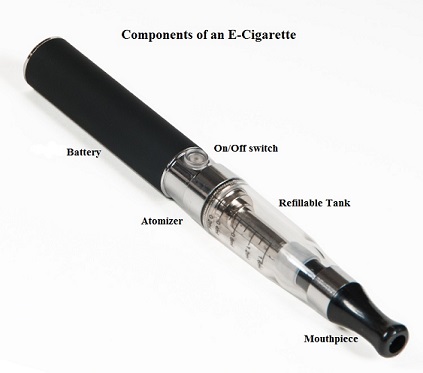
SUMMARY: The American Cancer Society estimates that tobacco use is responsible for nearly 1 in 5 deaths in the United States and accounts for at least 30% of all cancer deaths. Smokeless tobacco products are a major source of cancer causing nitrosamines, and increase the risk of developing cancer of the oropharynx, esophagus, and pancreas. Cigarette smoke contains more than 7,000 chemicals, many of which are toxic and some linked to cancer. E-cigarettes or Electronic Nicotine Delivery Systems (ENDS) were first developed in China and introduced to the U.S. market in 2007. When a smoker inhales through the mouth piece of an e-cigarette, the air flow triggers a sensor that switches on a small lithium battery powered heater, which in turn vaporizes liquid nicotine along with PolyEthylene Glycol (PEG) present in a small cartridge. The PEG vapor looks like smoke. The potent liquid form of nicotine extracted from tobacco is tinctured with fragrant flavors such as chocolate, cherry and bubble gum, coloring substances, as well other chemicals and these e-liquids are powerful neurotoxins.

With the rapid growth of the e-cigarette industry and the evidence of potential dangers and risk to public health, particularly children, experts from the world's leading lung organizations were compelled to release a position statement on electronic cigarettes, specifically focusing on their potential adverse effects on human health and calling on government organizations to ban or restrict the use of e-cigarettes, until their impact on health is better understood. According to the National Youth Tobacco Survey, the use of e-cigarettes has tripled from 2013 to 2014 among middle school and high school students. Epidemiological data have shown that nicotine use is a gateway to the use of cocaine and marijuana and subsequent lifelong addiction. E-cigarettes and other Electronic Nicotine Delivery Systems (ENDS), unlike combustible cigarettes and many other tobacco products are not currently regulated by the U.S. Food and Drug Administration.

Smoking more than one pack of cigarettes a day has been associated with an increased risk of mortality among patients with bladder cancer. E-cigarettes have been advertised and promoted as a safer way of delivering the stimulating effects of tobacco smoke, without the harmful health risks. Contrary to this claim, over 90% of inhaled nicotine is excreted to urine. Nicotine can be nitrosatized in urothelial cells and then further metabolized into carcinogenic nitrosamines and formaldehyde, which in turn can induce DNA damage in the bladder mucosa. Additionally, these metabolites can also block DNA repair, increasing cancer risk.

The authors in this study compared the urine of e-cigarette users to that of nonsmokers, for known bladder carcinogens. There are five known bladder carcinogens that are either present in traditional cigarettes or common solvents believed to be used in some e-cigarette formulations, and they include (benz(a)anthracene, benzo(a)pyrene, 1-hydroxypyrene, o-toluidine and 2-naphthylamine. The limit of detection of these carcinogens in this study was 10-100 ng/ml. Urine samples were collected from 13 e-cigarette users and 10 non-smoking, non e-cigarette using controls. The mean age was 39 years and participants were predominantly male and had abstained completely from traditional cigarettes, for at least 6 months prior to specimen collection.

It was noted that the urine from 92% of e-cigarette users tested positive for two of the five carcinogenic compounds whereas none of the controls tested positive for these carcinogens. Interestingly, all study participants considered e-cigarettes were safe, when interviewed by the study investigators.

It was concluded that in this important pilot study, majority of the e-cigarette users had carcinogenic metabolites in the urine, which can contribute to the development of bladder cancer. This study underscores the importance both traditional and e-cigarette smoking cessation, for bladder cancer prevention. MP88-14 EVALUATION OF E-CIGARETTES USERS URINE FOR KNOWN BLADDER CARCINOGENS. Fuller T, Acharya A, Bhaskar G, et al. DOI: http://dx.doi.org/10.1016/j.juro.2017.02.2739

Guideline for HER2 Testing in Gastroesophageal Adenocarcinoma

SUMMARY: It is estimated that in 2016 GastroEsophageal Adenocarcinoma (GEA) accounted for 43,280 new cases in the United States. Majority of the patients with GEA have advanced disease at the time of initial presentation and have limited therapeutic options with little or no chance for cure. Patients with localized disease (stage II and stage III) are often treated with multimodality therapy and 40% of the patients may survive for 5 years or more. Approximately 7-38% of GEAs have amplification and/or overexpression of HER2. HER2 (ERBB2) is a proto-oncogene located on the long arm of chromosome 17 (17q12) that encodes a tyrosine kinase receptor, which upon dimerization and phosphorylation, initiates signaling pathways, that lead to cell division, proliferation, differentiation and anti-apoptosis signaling.

GastroEsophageal junction tumors have a higher incidence of overexpression of HER2 compared to gastric cancers. In the stomach, overexpression of HER2 varies with histologic type (intestinal-type having greater expression than diffuse-type) and differentiation (well and moderately differentiated tumors having greater expression than poorly differentiated tumors). Further, the heterogeneity of immunostaining is greater in GEA and the complete membrane staining required for positivity in breast cancer, is not common in GEA. Due to the high heterogeneity of a gastric cancer, it is appropriate to evaluate HER2 expression on biopsy specimens, especially in unresectable cases and reevaluate it on resected specimens, when available.

The NCCN (National Comprehensive Cancer Network) Clinical Practice Guidelines in Oncology for Gastric Cancer and Esophageal and Esophagogastric Junction Cancers, recommend assessment of HER2 overexpression using ImmunoHistoChemistry and/or gene amplification using FISH (Fluorescence In Situ Hybridization or another in situ hybridization method, in tumor samples from patients, with unresectable, locally advanced, recurrent, or metastatic GEA, for whom HERCEPTIN® may be potentially beneficial. Testing for HER2 is primarily performed on Formalin-Fixed and Paraffin-Embedded biopsy or resection tumor tissue from the primary or metastatic site. There are important differences in HER2 expression, scoring, and outcomes in GEA compared to breast carcinoma, and the need for HER2 guidelines (that include critical clinical and laboratory considerations) was therefore recognized. The CAP, American Society for Clinical Pathology, and ASCO convened an international expert panel to systematically review published documents and to develop an evidence-based guideline, to establish recommendations for HER2 testing in GastroEsophageal Adenocarcinoma.

HERCEPTIN® (Trastuzumab) is humanized monoclonal antibody that targets HER2. HERCEPTIN® is effective only in cancers where HER2 is overexpressed. In a previously published randomized phase III trial, HERCEPTIN® in combination with chemotherapy was shown to significantly improve Overall Survival compared with chemotherapy alone, in patients with HER2 positive advanced GEA.

The following are the Guideline Summary from the College of American Pathologists, American Society for Clinical Pathology, and American Society of Clinical Oncology

Guideline Questions

• What is the optimal testing algorithm for the assessment of Human Epithelial Growth Factor 2 (HER2) status in patients with GastroEsophageal Adenocarcinoma (GEA)?

• What strategies can help ensure optimal performance, interpretation, and reporting of established assays in patients with GEA?

Target Population: Patients with GEA.

Target Audience: Medical and surgical oncologists; oncology nurses and physician assistants; pathologists; general practitioners; and patients.

Key Points and Recommendations for Clinicians

Recommendation 1.1

For patients with advanced GEA who are potential candidates for HER2-targeted therapy, the treating clinician should request HER2 testing on tumor tissue.

Recommendation 1.2

Treating clinicians or pathologists should request HER2 testing on tumor tissue in the biopsy or resection specimens (primary or metastasis) preferably before the initiation of HERCEPTIN® therapy if such specimens are available and adequate. HER2 testing on fine-needle aspiration specimens (cell blocks) is an acceptable alternative.

Recommendation 1.3

Treating clinicians should offer combination chemotherapy and HER2-targeted therapy as the initial treatment of appropriate patients with HER2-positive tumors who have advanced GEA

Key Points and Recommendations for Pathologists

Recommendation 2.1

Laboratories/pathologists must specify the antibodies and probes used for the test and ensure that assays are appropriately validated for HER2 ImmunoHistoChemistry (IHC) and in situ hybridization (ISH) on GEA specimens.

Recommendation 2.2

When GEA HER2 status is being evaluated, laboratories/pathologists should perform/order IHC testing first, followed by ISH (In Situ Hybridization) when the IHC result is 2+ (equivocal). Positive (3+) or negative (0 or 1+) HER2 IHC results do not require further ISH testing.

Recommendation 2.3

Pathologists should use the Ruschoff/Hofmann method in scoring HER2 IHC and ISH results for GEA.

Recommendation 2.4

Pathologists should select the tissue block with the areas of lowest grade tumor morphology in biopsy and resection specimens. More than one tissue block may be selected if different morphologic patterns are present.

Recommendation 2.5

Laboratories should report HER2 test results in GEA specimens in accordance with the College of American Pathologists biomarker Template for Reporting Results of HER2 (ERBB2) Biomarker Testing of Specimens From Patients With Adenocarcinoma of the Stomach or Esophagogastric Junction.

Recommendation 2.6

Pathologists should identify areas of invasive adenocarcinoma and also mark areas with strongest intensity of HER2 expression by IHC in GEA specimens for subsequent ISH scoring when required.

Recommendation 2.7

Laboratories must incorporate GEA HER2 testing methods into their overall laboratory quality improvement program, establishing appropriate quality improvement monitors as needed to ensure consistent performance in all steps of the testing and reporting process. In particular, laboratories performing GEA HER2 testing should participate in a formal proficiency testing program, if available, or an alternative proficiency assurance activity.

Recommendation 2.8

There is insufficient evidence to recommend for or against genomic testing in patients with GEA at this time.

HER2 Testing and Clinical Decision Making in Gastroesophageal Adenocarcinoma: Guideline Summary From the College of American Pathologists, American Society for Clinical Pathology, and American Society of Clinical Oncology. Bartley AN, Washington MK, Ismaila N, et al. Journal of Oncology Practice 2017;13:53-57.