Late Breaking Abstract – ASCO 2016 Liquid Biopsy Can Rapidly Detect Certain Gene Mutations with High Specificity

Late Breaking Abstract – ASCO 2016: Liquid Biopsy Can Rapidly Detect Certain Gene Mutations with High Specificity

SUMMARY: The FDA approved the first “Liquid Biopsy” test on June 1, 2016 for the detection of exon 19 deletions or exon 21 (L858R) substitution mutations in the Epidermal Growth Factor Receptor (EGFR) gene. On the heels of this approval, Zill and colleagues reported the results of the largest liquid biopsy study ever conducted thus far. It has been well established that treatment with EGFR TKIs results in superior outcomes, for patients with tumors harboring exon 19 deletions and exon 21 mutations. The application of precision medicine with targeted therapy requires detection of molecular abnormalities in a tumor specimen, following progression or recurrence. Archived biopsy specimens may not be helpful, as it is important to identify additional mutations in the tumor at the time of recurrence or progression, in order to plan appropriate therapy. Further, recurrent tumors may be inaccessible for a safe biopsy procedure or the clinical condition of the patient may not permit a repeat biopsy. Additionally, the biopsy itself may be subject to sampling error due to tumor heterogeneity. Genotyping cell free DNA in the plasma, also called liquid biopsy, can potentially overcome the shortcomings of repeat biopsies and tissue genotyping, allowing the detection of many more targetable gene mutations, thus resulting in better evaluation of the tumor genome landscape.
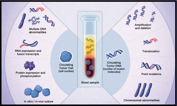
The authors in this study utilized Next Generation Sequencing (NGS) of circulating tumor DNA (ctDNA), isolated from plasma specimens (liquid biopsy specimens) of 15,191 patients of whom 37% had advanced lung cancer, 14% had breast cancer, 10% had colorectal cancer and 39% had other malignancies. Seventy genes were targeted and accuracy of ctDNA sequencing was assessed by comparing with matched tissue tests for 386 patients and frequencies of somatic ctDNA alterations per gene were compared to those previously described in tissue sequencing projects such as data from The Cancer Genome Atlas (TCGA).

It was noted that the ctDNA mutation patterns were highly concordant with tissue analysis as reported by the TCGA. The overall accuracy of ctDNA sequencing in comparison with matched tissue tests was 87% and the accuracy increased to 98% when blood and tumor were collected less than six months apart. Pearson Correlation between sets of data is a measure of how well these sets are related. Between 0.5 and 1.0 is considered high correlation. Pearson correlation for TP53 gene was 0.94, for KRAS was 0.99 and for PIK3CA was 0.99.

The researchers commented on the clinical outcome benefits using liquid biopsy, in four distinct groups:

1) Testing for actionable mutations (ALK fusion, EGFR or BRAF activating mutations in lung; ERBB2 amplification in gastric cancer) in cases with insufficient tissue quantity.

2) Testing for actionable resistance mutations (MET amplification or EGFR T790M in lung cancer), at the time of progression.

3) Genomic evolution upon progression such as ERBB2-amplified metastatic breast cancer in patients with triple negative primary tumor.

4) Tumors with genotypes that need more extensive driver mutation testing such as BRAF V600E in lung.

The authors concluded that there is a high correlation between ctDNA plasma samples and tissue testing with the exception of resistance mutations such as EGFR T790M mutation which evolve while on anti-EGFR inhibitor therapy and consequently may not correlate with the TCGA, probably because patients in the tissue-based population had not yet received the anti-EGFR inhibitor therapy that promotes the mutation. Patients who received treatment based on ctDNA findings also experienced better clinical outcomes. Zill OA, Mortimer S, Banks KC, et al Somatic genomic landscape of over 15,000 patients with advanced-stage cancer from clinical next-generation sequencing analysis of circulating tumor DNA. J Clin Oncol. 2016;34(suppl; abstr LBA11501).

Cancer Death Rate Declines in the US

SUMMARY: The American Cancer Society released the Cancer Statistics 2016 report, which includes the most recent data on cancer incidence, mortality, and survival in the US. It is estimated that in 2016, 1,685,210 new cancer cases will be diagnosed in the US and 595,690 cancer deaths are projected.

With a considerable decline in mortality from heart disease, cancer is now the leading cause of death in 21 states. In males, prostate cancer will be the leading cancer diagnosis in 2016 (21%) and Breast Cancer will be the leading cancer diagnosis in women (29%).

Lung Cancer remains the leading cause of cancer death both in men and women (27%). With major therapeutic advances against leukemia, brain cancer is now the leading cause of cancer death among children and adolescents (birth-19 years).

The overall cancer incidence rate in women has remained stable since 1998. However in men, cancer incidence has decreased by 3.1% per year since 2009 and this has been attributed to decline in routine screening with the PSA test. Routine screening with the PSA test is no longer recommended because of high rates of overdiagnosis, estimated at 23% to 42% for screen-detected cancers, which may not result in bad outcomes.

The cancer death rate in the US has dropped by 23% since 1991 which translates to more than 1.7 million deaths averted through 2012. There has been a continued decrease in death rates for the four major cancer sites – lung, breast, prostate, and colon/rectum. This overall decline in cancer deaths may be the result of reduction in smoking prevalence, improved screening modalities for breast, colon and prostate cancers and improvements in treatment.

Despite the overall reduction in cancer mortality, death rates are increasing for cancers of the liver, pancreas, and uterine corpus. Obesity has been shown to increase endometrial cancer risk by 50% for every 5 body mass index (BMI) units. Chronic infection with Helicobacter pylori and Hepatitis B virus has increased the incidence and death rates of stomach and liver cancer, respectively.

The authors concluded that “Advancing the fight against cancer will require continued clinical and basic research, which is dependent on funding, as well as the application of existing cancer control knowledge across all segments of the population, with an emphasis on disadvantaged groups.” With progress being made in cancer prevention using improved screening techniques and behavioral interventions, as well as rapid advances in cancer treatment with the understanding of cancer biology, it is expected that cancer death rate will continue to decline in the years to come. Cancer statistics, 2016. Siegel RL, Miller KD and Jemal A. CA Cancer J Clin 2016;66:7-30.

FDA Approves VISTOGARD®, an Antidote for 5-FU Overexposure

SUMMARY: The United States FDA approved VISTOGARD® (Uridine Triacetate) for the emergency treatment of adult and pediatric patients, who had severe or life-threatening toxicities within 4 days of treatment, following an overdose of 5-FluoroUracil (5-FU) or XELODA® (Capecitabine). VISTOGARD® is a Pyrimidine analog and following oral administration is deacetylated by nonspecific esterases, yielding Uridine in the circulation. Uridine is a direct antagonist of 5-FU and competitively inhibits 5-FU from incorporating in normal tissues, thus reducing cell damage and cell death.

The approval of VISTOGARD® was based on two separate trials in which 135 adult and pediatric cancer patients at increased risk for toxicity with 5-FU or XELODA® were included. Risk for toxicity could be due to 5-FU overdose and accidental XELODA® ingestion (N=111) or DihydroPyrimidine Dehydrogenase (DPD) deficiency and/or patients who experienced rapid onset of severe toxicities (N=24). These patients received VISTOGARD® granules 10 grams every 6 hours for 20 doses, starting within 96 hours after the termination of 5-FU therapy. The primary endpoint of the studies was survival at 30 days or until chemotherapy could resume, if prior to 30 days.

Of those who were treated with VISTOGARD® for overdose, 97 percent were still alive at 30 days. Of those treated with VISTOGARD® for early-onset severe or life-threatening toxicity, 89 percent were alive at 30 days. In both studies, 33 percent of patients resumed chemotherapy in less than 30 days. Adverse events were mild and uncommon and included nausea, vomiting and diarrhea.

The authors concluded that VISTOGARD® is a safe and effective antidote for 5-FU overexposure, and can facilitate rapid recovery and resumption of chemotherapy. Patients should take VISTOGARD® as soon as possible after overdose, regardless of symptoms or within 4 days of severe or life threatening toxicity. It should be noted that VISTOGARD® is not recommended for treatment of non-emergency adverse events associated with 5-FU and XELODA®, as this therapy may significantly decrease the efficacy of these chemotherapy agents. Clinical trial experience with uridine triacetate for 5-fluorouracil toxicity. Ma WW, Saif WM, El-Rayes BF, et al. J Clin Oncol 34, 2016 (suppl 4S; abstr 655)

Unique Toxicities of Immunotherapy for the Practicing Physician

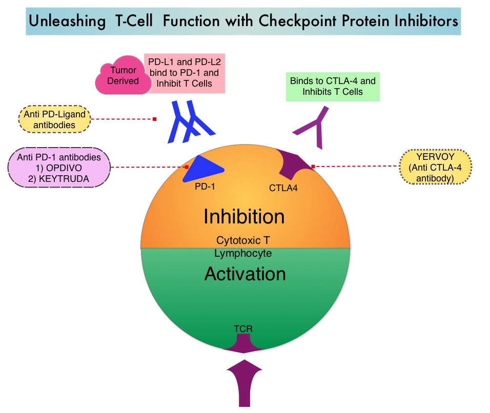
SUMMARY: Immunotherapy in cancer management includes Cancer Vaccines, Cytokine therapy, Adoptive Cell therapy and therapy with Check Point protein inhibitors such as YERVOY®, KEYTRUDA® and OPDIVO®. Toxicities related to these immunotherapeutic interventions are mediated by T cells resulting in exaggerated T cell response and potential damage to normal tissues. A brief summary of the more common adverse events associated with cancer immunotherapy, is listed below-

CANCER VACCINES

PROVENGE® (Sipuleucel-T) is an autologous, cellular immunotherapy indicated for the treatment of asymptomatic or minimally symptomatic metastatic Castrate Resistant (hormone-refractory) Prostate Cancer. This product is the only currently approved Cancer Vaccine and consists of autologous CD54+ cells activated with recombinant PAP/GM-CSF (Prostate Acid Phosphatase, an antigen expressed in the prostate cancer tissue, linked to immune cell activator, Granulocyte Macrophage-Colony Stimulating Factor). Vaccine therapies work by promoting type 1 or type 2 immune reactions. In type 1 immune reaction, T helper type 1 (Th1) lymphocytes secrete Interleukin-2 (IL-2), Interferon gamma, and lymphotoxin-alpha and facilitate intense phagocytic activity whereas in type 2 immunity, Th2 cells secrete IL-4, IL-5, IL-9, IL-10, and IL-13 and is characterized by high antibody titers. Cancer Vaccines are associated with minimal toxicities because the antigens associated with the tumor are overexpressed in the cancer cells and are not usually detectable in normal cells. Common side effects include local reactions, fever, chills, fatigue, rash, back pain and Melanoma vaccines are associated with vitiligo.

CYTOKINE THERAPY

Both INTRON® A (Interferon alfa-2b) and ROFERON® A (Interferon alfa-2a) are approved for a variety of malignant conditions as well as for Chronic Hepatitis B and C. In addition to fever, chills and flu like symptoms, two thirds of the patients have nausea and anorexia and up to 45% of the patients may experience symptoms of depression. Patients should be monitored for cytopenias, diarrhea, liver toxicities as well as thyroid dysfunction and autoimmune disorders may be exacerbated with Interferon.

PROLEUKIN® (High dose IL-2) is administered in an inpatient setting with cardiac monitoring, as patients often develop capillary leak syndrome and hypotension in addition to flu like symptoms and liver function abnormalities. This has been attributed to release of Nitric Oxide, IL-1, Tumor Necrosis Factor alpha, and IFN gamma. Patients may also develop autoimmune related thyroid dysfunction, cytopenias as well as neurotoxicity and will therefore require close monitoring.

ADOPTIVE CELL THERAPY

Unlike Cancer Vaccines, Adoptive T cell therapy is a type of passive immunization which involves the transfusion of autologous or allogeneic T cells into patients with malignancies. These tumor reactive T cells can be genetically engineered or grown ex vivo and their efficacy can be enhanced by other immunotherapies, such as Cancer Vaccines, Cytokine administration or in some instances cytotoxic chemotherapy and radiation therapy. BLINCYTO® (Blinatumomab) is a genetically engineered bispecific CD19 directed CD3 T-cell engager, approved by the FDA, that binds to CD19 (expressed on B-cells) and CD3 (expressed on T-cells). It is indicated for the treatment of Philadelphia chromosome-negative relapsed or refractory B-cell precursor Acute Lymphoblastic Leukemia (ALL). Administration of BLINCYTO® or high dose IL-2 given along with T cells, can cause Cytokine Release Syndrome (CRS), associated with fever, tachycardia, vascular leak, oliguria, and hypotension. This has been attributed to IL-6 and ACTEMRA® (Tocilizumab), an IL-6 receptor antagonist may be of benefit for these patients along with IV fluids, nonsteroidal anti-inflammatory agents and vasopressors. Other toxicities that require monitoring include flu like symptoms, liver function abnormalities, B-cell aplasia, cytopenias and neurotoxicity.

THERAPY WITH CHECKPOINT INHIBITORS

The FDA approved checkpoint inhibitors include, YERVOY® (Ipilimumab) which targets CTLA-4, KEYTRUDA® (Pembrolizumab) and OPDIVO® (Nivolumab), which block checkpoint PD-1. The toxicities associated with YERVOY® are dose dependant. Some common side effects of check point inhibitors include skin rash, flu like symptoms, liver function abnormalities, diarrhea and colitis, cytopenias, thyroid and adrenal function abnormalities. Rare cases of pneumonitis, encephalitis, Guillain-Barré syndrome, and a myasthenia gravis–like syndrome have been reported. With close monitoring, early diagnosis and intervention with Corticosteroids, these toxicities can be alleviated. REMICADE® (Infliximab), a chimeric monoclonal antibody against Tumor Necrosis Factor alpha (TNF-alpha), should be offered to those whose colitis does not resolve within 3 days of high dose steroids or for relapse of colitis with steroid taper.

Toxicities of Immunotherapy for the Practitioner. Weber JS, Yang JC, Atkins MB, et al. J Clin Oncol 2015;33:2092-2099

VARUBI® Now Approved for Delayed Chemotherapy Induced Nausea and Vomiting

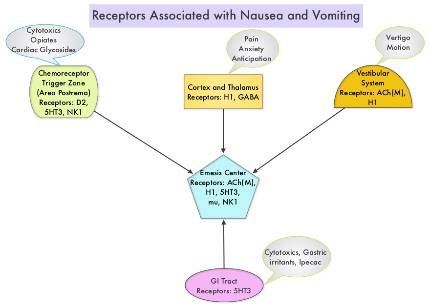
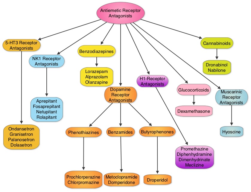
SUMMARY: The U.S. Food and Drug Administration on September 2, 2015, approved VARUBI® (Rolapitant) to prevent delayed phase Chemotherapy Induced Nausea and Vomiting (CINV). Chemotherapy Induced Nausea and Vomiting (CINV) is one of the most common adverse effects of chemotherapy and is experienced by about 80% of patients receiving chemotherapy. The development of effective antiemetic agents has facilitated the administration of majority of the chemotherapy agents in an outpatient setting avoiding hospitalization. Acute CINV begins within the first 24 hours following chemotherapy administration, with most patients experiencing symptoms within the first four hours of treatment, whereas delayed nausea and vomiting occurs more than 24 hours after chemotherapy administration and can persist for several days. Delayed CINV is often underestimated and a third of the patients receiving chemotherapy may experience delayed nausea and vomiting without prior acute nausea or vomiting. Acute nausea and vomiting is dependent on Serotonin (5-hydroxytryptamine-5HT3) and its receptors, with the chemotherapeutic agents stimulating the release of Serotonin from the enterochromaffin cells of the small intestine. 5-HT3 receptors are located on vagal afferent pathway, which in turn activates the vomiting center to initiate the vomiting reflex. 5-HT3 receptors are also located centrally in the Chemoreceptor Trigger Zone of the area Postrema. Delayed nausea and vomiting is associated with the activation of Neurokinin 1 (NK1) receptors by substance P. NK1 receptors are broadly distributed in the central and peripheral nervous systems. VARUBI® is a substance P/Neurokinin-1 (NK-1) receptor antagonist.

The safety and efficacy of VARUBI® were established in three randomized, double-blind, controlled clinical trials where VARUBI® in combination with KYTRIL® (Granisetron) and Dexamethasone was compared with placebo, KYTRIL® and Dexamethasone (control therapy), in more than 2500 patients receiving a moderately or highly emetic chemotherapy regimen. HEC Study 1 and HEC Study 2 included Cisplatin Based Highly Emetogenic Chemotherapy (HEC). Chemotherapy regimens included more than 60 mg/m2 of Cisplatin. In HEC Study 1, 532 patients were randomized to receive either antiemetic regimen with VARUBI® (N =266) or control therapy (N =266). In HEC Study 2, a total of 555 patients were randomized to receive either antiemetic regimen with VARUBI® (N =278) or control therapy (N =277). MEC Study 3 included Moderately Emetogenic Chemotherapy and combinations of Anthracycline and Cyclophosphamide chemotherapy. A total of 1369 patients were randomized in this study to receive either antiemetic regimen with VARUBI® (N =684) or control therapy (N =685). Patients in these trials received either VARUBI® 180 mg PO or placebo at 1 to 2 hours before administration of Highly Emetogenic Chemotherapy. All patients received intravenous KYTRIL® 10 μg/kg IV and Dexamethasone 20 mg PO on day 1 and Dexamethasone 8 mg PO twice daily on days 2 to 4 for up to six cycles, with each cycle lasting a minimum of 14 days. The primary endpoint in all three studies was complete response (defined as no emetic episodes and no rescue medication) in the delayed phase (25 to 120 hours) post chemotherapy.

It was noted that a significantly greater proportion of patients receiving antiemetic regimen with VARUBI® had complete responses in the delayed phase than did patients in the control therapy group – HEC Study 1: 72.7% vs 58.4% (P<0.001), HEC Study 2: 70.1% vs 61.9% (P=0.043) and MEC Study 3: 71.3% vs 61.6% (P<0.001). The most common adverse events in patients treated with VARUBI® included neutropenia, hiccups, decreased appetite and dizziness. It was concluded from these three trials that VARUBI® when combined with a 5-HT3 receptor antagonist such as KYTRIL® and a corticosteroid, significantly prevented delayed Chemotherapy Induced Nausea and Vomiting.

1) Safety and efficacy of rolapitant for prevention of chemotherapy-induced nausea and vomiting after administration of cisplatin-based highly emetogenic chemotherapy in patients with cancer: two randomised, active-controlled, double-blind, phase 3 trials. Rapoport BL, Chasen MR, Gridelli C, et al. The Lancet Oncology 2015;16:1079-1089

2) Phase 3 trial results for rolapitant, a novel NK-1 receptor antagonist, in the prevention of chemotherapy-induced nausea and vomiting (CINV) in subjects receiving moderately emetogenic chemotherapy (MEC). Schnadig ID, Modiano MR, Poma A, et al. J Clin Oncol 32:5s, 2014 (suppl; abstr 9633)

Hepatitis B Antiviral Prophylaxis for Cancer Patients with Solid Tumors Receiving Chemotherapy

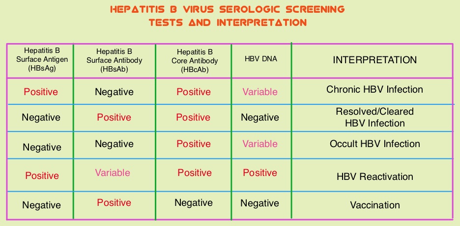
SUMMARY: The Centers for Disease Control and Prevention (CDC) estimates that there are 800,000 -1.4 million individuals with Chronic Hepatitis B (HBV) infection in the United States. Reactivation of HBV is a major concern in cancer patients who may be on chemotherapy or other immunosuppressive therapies, with the incidence of HBV reactivation ranging from 40%-60% in those who are positive for Hepatitis B surface antigen (HBsAg). HBV reactivation is preventable with prophylactic antiviral therapy, failing which it can result in delays in cancer treatment, as well as potentially fatal outcomes. Based on recently published data, showing the high risk for HBV reactivation among patients with hematological malignancies receiving B-cell-depleting agents such as RITUXAN® (Rituximab) or ARZERRA® (Ofatumumab), the FDA has urged health care providers to screen all patients for HBV infection, prior to starting therapy with these agents. HBV reactivation has been observed following chemotherapy for solid tumors, but the risk for reactivation in these settings has been unclear with insufficient evidence. The American Society of Clinical Oncology in 2010 rendered a Provisional Clinical Opinion (PCO), suggesting that there was insufficient evidence to determine the net benefits and harms of routine screening for HBV infection, in patients receiving chemotherapy and the recommendation was that screening be considered in those at increased risk for HBV infection or who receive highly immunosuppressive regimens.

This present study was conducted to determine the risk for HBV reactivation with and without antiviral prophylaxis and the benefit of prophylaxis in adults with solid tumors and chronic or resolved HBV infection. This meta-analysis included 26 original reports and the studies were independently reviewed by two investigators for study inclusion. HBV patients included in this study were receiving chemotherapy for any solid tumor with or without concomitant HBV prophylactic therapy. Study patients could receive long-term antiviral treatment or prophylaxis before chemotherapy initiation and the comparison was with those receiving chemotherapy without antiviral prophylaxis. The primary outcome was HBV reactivation as defined by a greater than 10-fold increase in HBV DNA levels from baseline or an absolute increase greater than 105 copies/mL in those with chronic HBV infection or the re-emergence of HBsAg when previously negative, in those with resolved HBV infection. Secondary outcomes included HBV-related hepatitis, interruption or delay in chemotherapy, acute liver failure with coagulopathy and hepatic encephalopathy and death.

It was noted that in patients with chronic HBV infection receiving chemotherapy, the risk for HBV reactivation without antiviral prophylaxis ranged from 4% to 68% (median, 25%). The risk for HBV reactivation, HBV-related hepatitis, and chemotherapy interruption was reduced by more than 80% with antiviral prophylaxis. Interestingly, in patients with resolved HBV infection receiving chemotherapy, there was still a risk of HBV reactivation, with this risk ranging from 0.3% to 9%. The authors in this meta-analysis addressed a very important question and concluded that the risk for HBV reactivation in patients with chronic HBV, on chemotherapy for solid tumors, is similar to the risk with other types of immunosuppressive therapy. Cancer patients should therefore be screened for HBV before chemotherapy is initiated for solid tumors and started on antiviral prophylaxis. Paul S, Saxena A, Terrin N, et al. Hepatitis B Virus Reactivation and Prophylaxis During Solid Tumor Chemotherapy: A Systematic Review and Meta-analysis. Ann Intern Med. 2016; 164:30-40.

The 2015 ASH CHOOSING WISELY® Campaign Five Hematologic Tests and Treatments to Question

SUMMARY: CHOOSING WISELY® is a quality improvement initiative led by the American Board of Internal Medicine Foundation in collaboration with leading medical societies in the United States such as the American Society of Hematology (ASH). This organization was established to improve quality of medical care, after it was noted that about 25% of the tests ordered at the time of hospital admission and 65% of the tests ordered on subsequent days were avoidable. Further, there is ample evidence to suggest that, reducing unneeded investigations can decrease costs, increase patient satisfaction and quality of care. CHOOSING WISELY® has challenged 70 medical societies to identify 5 tests, procedures or treatments, within each specialty’s clinical domain, that are offered to patients, despite the lack of evidence demonstrating its benefit. The goal is to make positive changes in the actual delivery of patient care without harming the patient. The ASH CHOOSING WISELY® Task Force comprised of 13 individuals, represents a broad spectrum of hematologic expertise including malignant, benign, adult, and pediatric specialists. The five final recommendations of the 2015 ASH Choosing Wisely Campaign is an addition to the 10 prior recommendations made by ASH over the past 2 years. These top 5 recommendations were presented on December 7, 2015, at the 57th annual meeting of ASH, in Orlando, Florida. Practicing hematologists should give due consideration to these recommendations which are evidence based and cost effective.

Don’t image for suspected Pulmonary Embolism (PE) without moderate or high pre-test probability of PE

The American College of Radiology has recommended that assessment of the risk-benefit ratio is important especially with pulmonary embolism and imaging can be avoided for suspected PE, without moderate to high pre-test probability.

Don’t routinely order thrombophilia testing on patients undergoing a routine infertility evaluation

With Nearly 15% couples of patients receiving an infertility evaluation, the American Society for Reproductive Medicine has recommended that even though several population-based studies have found association of infertility or failure of assisted reproduction with thrombophilia, 2 large cohort studies have shown no association between thrombophilias such as Factor V Leiden or Prothrombin gene mutations and assisted reproduction failure or infertility. Further, thrombophilia is not a predictor of who will benefit from Low Molecular Weight Heparin (LMWH) treatment with respect to assisted reproduction and LMWH can be associated with adverse events.

Don’t perform repetitive Complete Blood Count (CBC) and chemistry testing in the face of clinical and lab stability

The Society for Hospital Medicine and Adult Hospital Medicine noted that ordering routine complete blood counts (CBCs) during hospitalization is common practice and is unnecessary. Critically ill patients do not have the bone marrow reserve or erythropoietin stimulus to compensate for iatrogenic blood loss. Reducing the frequency of CBC’s does not result in inferior outcomes and several studies have shown that there is no difference in readmission rates, length of hospital stay and rates of adverse events. In addition to the risks of phlebotomy, this practice is economically disadvantageous, as they may not be reimbursable and will be an additional avoidable cost to dispose the biohazard waste of the blood samples.

Don’t transfuse red blood cells for iron deficiency without hemodynamic instability

The American Association of Blood Banks has recommended against PRBC transfusions for patients with hemodynamically stable iron deficiency anemia. These patients when evaluated in the Emergency Department (ED) can be prescribed oral or IV iron with similar responses noted at 6-8 weeks. The compliance rate in those receiving oral iron may only be 50% due to GI side effects. Therefore parenteral iron may be a better treatment option for certain groups of patients seen in the ED.

Avoid using positron emission tomography (PET) or PET-CT scanning as part of routine follow-up care to monitor for a cancer recurrence in asymptomatic patients who have finished initial treatment to eliminate the cancer unless there is high-level evidence that such imaging will change the outcome

Professional organizations like ASCO, ESMO and NCCN do not include surveillance PET in disease-specific guidelines because, routine use of intensive surveillance does not improve survival or enhance quality of life. Besides cost implications, CT scans may in fact expose patients to small doses of radiation.

The ASH Choosing Wisely® Campaign: Top 5 Non-ASH Choosing Wisely® Recommendations of Relevance to Hematology. Presented on December 7, 2015, at the 57th annual meeting of ASH, in Orlando, Florida.

ZOMETA® Administered Every 12 Weeks Is Non-inferior to Every 4 Weeks for Bone Metastases

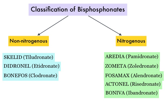
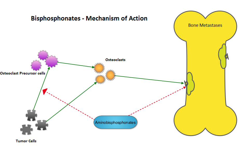
SUMMARY: Bones are the third most common site of metastatic disease and approximately 100,000 cases of bone metastasis are reported in the United States each year. Cancers originating in the breast, prostate, lung, thyroid and kidney, are more likely to metastasize to the bone. Bisphosphonates inhibit osteoclast-mediated bone resorption and both oral and IV bisphosphonates reduce the risk of developing Skeletal Related Events (SRE’s) and delay the time to SRE’s in patients with bone metastases. Bisphosphonates can also reduce bone pain and may improve Quality of life. Intravenous bisphosphonates, Pamidronate (AREDIA®) and Zoledronic acid (ZOMETA®) have been approved in the US for the treatment of bone metastases. Amino-bisphosphonate, ZOMETA® has however largely replaced AREDIA®, because of its superior efficacy. Both AREDIA® and ZOMETA® are administered IV every 3 to 4 weeks during the first year, following diagnoses of bone metastases. However, the optimal treatment schedule following this initial phase of treatment has remained unclear. Further, renal toxicity, long bone fractures and OsteoNecrosis of the Jaw (ONJ) have been identified as potential problems with bisphosphonate use.
CALGB 70604 (Alliance), is a randomized phase III study in which the efficacy of ZOMETA® administered every 4 weeks was compared with ZOMETA® administered every 12 weeks, in patients with breast cancer, prostate cancer or multiple myeloma, with bone metastases. In this non-inferiority trial, 1822 patients (Breast = 833, Prostate = 674, Myeloma= 270 and Other= 45) were randomly assigned 1:1, to receive ZOMETA® every 4 weeks or every 12 weeks for 2 years. The primary endpoint was incidence of any Skeletal Related Event (SRE) and secondary endpoints included skeletal morbidity rates, performance status, pain using the Brief Pain Inventory and incidences of ONJ and renal dysfunction. Both treatment groups were well matched. Patients in this trial were stratified by disease and analyses by disease was pre-planned. It was noted that for the primary endpoint, there was no significant difference between the two treatment groups with 29% of patients in both treatment groups experiencing at least one SRE (P=0.79). With regards to secondary endpoints, there were still no significant differences between the two treatment groups, including renal dysfunction and ONJ. The authors pointed out that toxicities such as ONJ and renal dysfunction are more likely to occur after 2 years of treatment.
It was concluded that ZOMETA® administered every 3 months for 2 years is non-inferior to ZOMETA® administered every 4 weeks for 2 years, in patients with breast cancer, prostate cancer and multiple myeloma, with bone metastases. A less frequent dosing of ZOMETA® compared with the standard monthly dosing, may be more convenient for the patients and cost effective. CALGB 70604 (Alliance): A randomized phase III study of standard dosing vs. longer interval dosing of zoledronic acid in metastatic cancer. Himelstein AL, Qin R, Novotny PJ, et al. J Clin Oncol 33, 2015 (suppl; abstr 9501)

FDA Approves First Biosimilar Product ZARXIO® – A Primer on Biosimilars

SUMMARY: The U.S. FDA on March 6, 2015 approved ZARXIO® (Filgrastim-sndz), the first biosimilar product approved in the United States. A biosimilar product is a biological product that is approved based on its high similarity to an already approved biological product (also known as reference product). Biological products are made from living organisms including humans, animals and microorganisms such as bacteria or yeast and are manufactured through biotechnology, derived from natural sources or produced synthetically. Biological products have larger molecules with a complex structure than conventional drugs (also known as small molecule drugs). Unlike biological products, conventional drugs are made of pure chemical substances and their structures can be identified. A generic drug is a copy of brand name drug and has the same active ingredient and is the same as brand name drug in dosage form, safety and strength, route of administration, quality, performance characteristics and intended use. Therefore, brand name and the generic drugs are bioequivalent.

The Affordable Care Act in 2010 created an abbreviated licensure pathway for biological products that are demonstrated to be “biosimilar” to, or “interchangeable” with an FDA-licensed (FDA approved) biological product (reference product). The biosimilar must show that it has no clinically meaningful differences in terms of safety and effectiveness from the reference product. A biosimilar product can only be approved by the FDA if it has the same mechanism of action, route of administration, dosage form and strength as the reference product, and only for the indications and conditions of use that have been approved for the reference product. Biosimilars are not as easy to manufacture as generics (copies of brand name drugs) because of the complexity of the structure of the biologic product and the process used to make a biologic product. The facilities where biosimilars are manufactured must also meet the FDA’s standards.

The FDA’s approval of ZARXIO® was based on review of evidence that included structural and functional characterization, animal study data, human pharmacokinetic and pharmacodynamics data, clinical immunogenicity data and other clinical safety and effectiveness data, that demonstrated ZARXIO® was biosimilar to NEUPOGEN®. ZARXIO® was approved as a biosimilar and not as an interchangeable product (Can only be substituted for the reference product after approval by the prescribing Health Care Provider). ZARXIO® is approved for the same indications as NEUPOGEN® and these indications include

• Patients with cancer receiving myelosuppressive chemotherapy

• Patients with Acute Myeloid Leukemia receiving induction or consolidation chemotherapy

• Patients with cancer undergoing Bone Marrow Transplantation

• Patients undergoing Autologous peripheral blood progenitor cell collection and therapy

• Patients with severe Chronic Neutropenia.

The most common expected side effects of ZARXIO® are bone and muscle aches, redness, swelling or itching at injection site. Less common, serious side effects include spleen rupture and serious allergic reactions. Unlike ZARXIO® which was approved via an abbreviated licensure pathway for biosimilars, GRANIX® (tbo-Filgrastim) was approved via the full Biologic License Application pathway, which presently limits GRANIX® use only for reducing the duration of severe neutropenia in patients non-myeloid malignancies, receiving myelosuppressive chemotherapy. The present Medicare reimbursement rules will be more favorable to ZARXIO® compared to GRANIX®, based on their approval process. FDA approves first biosimilar product ZARXIO®. http://www.fda.gov/NewsEvents/Newsroom/PressAnnouncements/ucm436648.htm

Fish Oil and Certain Species of Fish May Negate the Effects of Chemotherapy

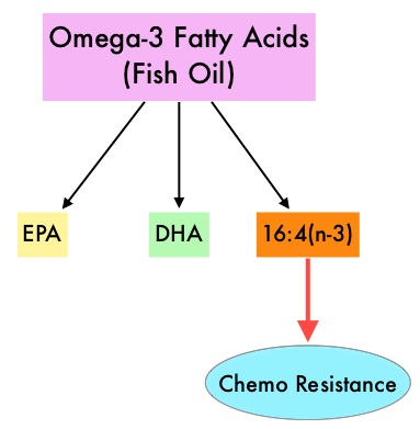
SUMMARY: It is estimated that approximately 20% of cancer patients in the US take Omega-3 fatty acids in the form of fish oil. Fish oil is a mixture of fatty acids produced from several species of fish and the two most abundant and important fatty acids in fish oil include EicosaPentaenoic Acid (EPA) and DocosaHexaenoic Acid (DHA). Fish oil content in presently available preparations is not standardized and does not require FDA approval. Preclinical studies have demonstrated that mouse tumors recruit mesenchymal stem cells that are specifically activated by platinum based chemotherapy and secrete 2 fatty acids, 12S-HHT and 16:4(n-3)). These fatty acids are called Platinum Induced Fatty Acids (PIFAs) and they have been shown to induce resistance to a broad range of chemotherapeutic agents, by activating a cytoprotective response in the tumor tissue. Fish oil has relevant levels of fatty acid 16:4(n-3) and preclinical models have shown that the fish oil neutralized the antitumor activity of chemotherapy, thus conferring drug resistance. With this preclinical information and given that cancer patients frequently use fish oil supplements, the authors evaluated the effect of fish oil intake in healthy volunteers, on the plasma levels of fatty acid 16:4(n-3), which has been shown to induce resistance to chemotherapeutic agents. The researchers first conducted a survey to determine what percentage of cancer patients undergoing treatment at a University Medical Center in the Netherlands were taking fish oil supplements. They also analyzed fatty acid 16:4(n-3) content, in 3 brands of fish oil supplements and 4 often consumed species of fish. The authors then randomly selected 30 healthy volunteers for the fish oil study and 20 healthy volunteers for the fish consumption study and the plasma levels of fatty acid 16:4(n-3) was measured after they consumed fish oil or fish, for a period of 2 weeks. They noted that 11% of the cancer patients in their study reported using omega-3 supplements. All fish oils tested contained amounts of fatty acid 16:4(n-3) ranging from 0.2 to 5.7 μM and this was adequate to induce chemoresistance to a variety of chemotherapeutic agents. They noted that there was a significant rise in the plasma 16:4(n-3) fatty acid levels in the healthy volunteers after they consumed fish oil supplements and fish, with high levels of fatty acid 16:4(n-3). Herring and Mackerel fish contained high levels of fatty acid 16:4(n-3), in contrast to Salmon and Tuna. The authors concluded that based on this preclinical data it is best to avoid fish oils and fish such as Herring and Mackerel in the 48 hours surrounding chemotherapy, as the high plasma 16:4(n-3) fatty acid levels may negate the effects of chemotherapy. These recommendations have been adopted by the Dutch Cancer Society and by the Dutch National Working Group for Oncologic Dieticians. Increased Plasma Levels of Chemoresistance-Inducing Fatty Acid 16:4(n-3) After Consumption of Fish and Fish Oil. Daenen LGM, Cirkel GA, Houthuijzen JM, et al. JAMA Oncol. 2015;1:350-358