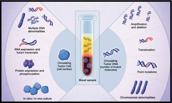
Late Breaking Abstract – ASTRO 2019 Detectable HPV Circulating Tumor DNA in Post-Operative Oropharyngeal Squamous Cell Carcinoma Patients is Associated with Progression

SUMMARY: The Centers for Disease Control and Prevention estimates that in the US, there are more than 16,000 cases of Human PapillomaVirus (HPV)-positive OroPharyngeal Squamous Cell Carcinoma (OPSCC) per year and there has been a significant increase in incidence during the past several decades. They represent approximately 70% of all OPSCC in the United States and Canada. HPV is a non-enveloped, double-stranded, DNA virus that infects epithelial cells and majority of the HPV subtypes cause epithelial lesions such as warts or papillomas, which are of low malignant potential. However, there is a subset of high-risk HPV that can cause cancer by integrating its DNA into the host genome, with the resulting expression of two important oncogenes E6 and E7 in the host cell. The E6 oncogene binds and degrades tumor suppressor TP53 via ubiquitin-mediated processes thereby preventing the host cell from engaging in cell cycle checkpoints and enduring an apoptotic response. The E7 oncogene binds to and destabilizes tumor suppressor retinoblastoma (pRb) resulting in transcription of genes involved in proliferation and cell cycle progression. One of the main molecular pathways amplified through E7 is the CDKN2A/p16 gene pathway, which results in the overexpression of p16 protein. E7 also induces cellular proliferation by disrupting the activity of Cyclin Dependent Kinase inhibitors p21 and p27. In essence, HPV infection induces failures in cell cycle checkpoints, resulting in genetic instability and over time, progression of premalignant lesions to invasive Squamous Cell Carcinoma. Unlike tobacco induced HNSCC where TP53 and pRb pathways are nullified due to mutation and epigenetic alterations, in HPV-related HNSCC, wild-type TP53 and pRb are functionally inactivated by the viral oncogenes.

Patients with HPV-positive OPSCC tend to be younger males, who are former smokers or nonsmokers, with risk factors for exposure to High Risk HPV infection. The HPV-positive primary Squamous Cell Carcinoma tends to be smaller in size, with early nodal metastases, and HPV status particularly in OroPharyngeal Squamous Cell Carcinoma is an independent prognostic factor for Overall Survival (OS) and Progression Free Survival (PFS). These patients have a better prognosis compared with patients with HPV-negative Head and Neck Squamous Cell Carcinoma (HNSCC), when treated similarly. Further, HPV positive patients demonstrate higher Response Rates to chemoradiation as well as an improved Overall Survival. However, approximately 20% of patients diagnosed with HPV-positive OPSCC experience cancer progression within 5 years.

The role of circulating tumor DNA (ctDNA) as a cancer biomarker in the post-operative surveillance of patients with HPV-associated OPSCC has remained unclear. The authors in this study aimed to investigate ctDNA detectability rates by post-op risk category and association with prognosis in this patient population. The researchers prospectively collected and tested serum samples from 29 patients with HPV-associated OPSCC who had not yet undergone treatment, for assay validation. As a control, 7 HPV negative OPSCC patients were included. A cohort of 46 patients with HPV-associated OPSCC who had undergone surgery for the disease had serum samples collected prior to beginning adjuvant therapy. The serum collected from this total group of 82 patients (N=29+7+46) was analyzed in a blinded fashion for E6/E7 HPV ctDNA using ddPCR multiplex assay (HPV 16, 18, 31, 33), and HPV ctDNA detectability was compared statistically across groups. Associations of patient and tumor characteristics with recurrence were assessed and estimates of Progression Free Survival (PFS) and Overall Survival (OS) were made using the Kaplan-Meier (KM) method.

The researchers found that ctDNA was detectable in 27 of 29 patients who had not yet undergone treatment, for a sensitivity rate of 93%, whereas none of the 7 HPV-negative patients had detectable ctDNA, for a specificity rate of 100%. Post-op serum was collected at a median of 25 days after surgery prior to beginning adjuvant therapy and ctDNA was detectable in 43% of patients including 47% with high-risk features (Extra Nodal Extension or R1-microscopic residual tumor). All detected ctDNA was HPV type 16.

At a median follow up of 20 months for the post-op HPV-OPSCC cohort, 24% of these patients had recurrent disease. Among those who recurred, 64% had detectable ctDNA compared with 35% whose disease did not recur (P=0.1). There was a significant association between detectable ctDNA and 24-month Progression Free Survival (45% versus 84%; P=0.04) and Overall Survival (80% versus 100%; P=0.02). Overall, detectable ctDNA, T4 tumors, and more than 4 positive lymph nodes were positively associated with disease recurrence.

It was concluded that detectable HPV circulating tumor DNA is a highly sensitive and specific means of determining HPV-status and was significantly associated with worsened Progression Free Survival and Overall Survival among post-op patients with Human Papilloma Virus (HPV)-associated OroPharyngeal Squamous Cell Carcinoma. HPV circulating tumor DNA as a cancer biomarker may also assist in risk stratification, treatment assessment, and surveillance. Detectable HPV ctDNA in Post-Operative Oropharyngeal Squamous Cell Carcinoma Patients is Associated with Progression. Routman DM, Chera BS, Jethwa KR, et al. International Journal of Radiation Oncology • Biology • Physics 2019;105, 682-683. LBA5

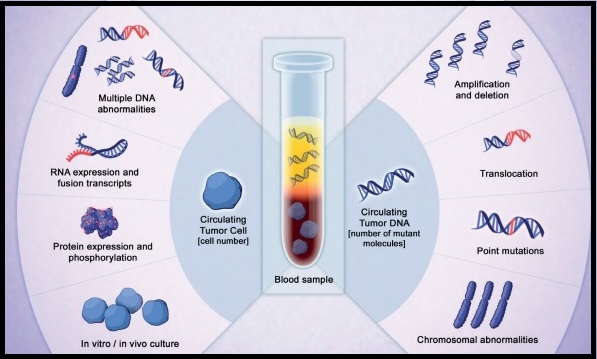
Clinical Utility of NETest®, a Liquid Biopsy Assay for Diagnosis, Monitoring Therapy and Prognosis, in Patients with Neuroendocrine Tumors

SUMMARY: It is estimated that in the United States, more than 12,000 people are diagnosed with a Neuroendocrine tumor each year. NeuroEndocrine Tumors (NETs) arise from cells of the endocrine and nervous systems and produce biogenic amines and polypeptide hormones. NETs can be clinically symptomatic (functioning) or silent (nonfunctioning). The incidence is higher in African-Americans and the most common sites of NETs are the lung, stomach, appendix, cecum, duodenum, pancreas, jejunum/ileum, colon, and rectum. NeuroEndocrine Tumors originating in the gastrointestinal tract and pancreas are also known as GastroEnteroPancreatic NETs (GEP-NETs). They constitute about 2% of all neoplasms and account for about 50-70% of all NETs. They are more frequent in gastric fundus/body, proximal duodenum, Vater’s papilla, pancreas, tip of the appendix, terminal ileum, and lower rectum. Majority of GEP-NETs are not symptomatic (nonfunctioning tumors), difficult to diagnose, and present with advanced disease at initial diagnosis. They often metastasize to the mesentery, peritoneum and liver. The functioning tumors however secrete biologically active substances that can lead to the development of characteristic clinical syndromes. NeuroEndocrine Tumors may be sporadic or may be a component of inherited genetic syndromes such as Multiple Endocrine Neoplasia (MEN) types 1 and 2. Most NETs are classified based on tumor differentiation into 1) Well-differentiated, Low-grade (G1) 2) Well-differentiated, Intermediate-grade (G2) and 3) Poorly differentiated, High-grade (G3). Tumor differentiation and tumor grade often correlate with mitotic count and Ki-67 proliferation index. Even though surgery is curative when the tumor is detected early, this is often not the case, as most patients present with metastatic disease at the time of diagnosis.

Chromogranin A is a glycoprotein precursor to several functional peptides and is considered a standard biomarker for NETs. However, serum Chromogranin A levels can be elevated in several non-oncologic conditions such as atrophic gastritis, pancreatitis, chronic hepatitis, liver cirrhosis, irritable bowel, and inflammatory bowel diseases. The use of proton pump inhibitors can also result in elevated Chromogranin A levels. Diagnostic imaging as well as serum biomarkers lack the sensitivity and are unable to detect early changes in disease state. As such, the absence of a clinically useful blood biomarker remains an important unmet need.

The NETest® is a novel blood-based (liquid biopsy) molecular diagnostic test intended to aid in the identification of neuroendocrine tumor disease activity. The assay involves measurement of 51 neuroendocrine tumor gene transcripts, by Polymerase Chain Reaction (PCR). The gene expression signatures, which is the tumor activity score, stratifies patients into three groups: low score (40% or less), moderate/intermediate score (41-79%) and high score (80% or more). A higher score at the time of testing indicates a higher risk of tumor activity. Previously published prospective clinical studies have demonstrated the value of NETest® in predicting the effectiveness of surgery, in its ability to monitor tumor progression during SomatoStatin Analog (SSA) therapy, in its utility for watch‐and‐wait programs, as well as its ability in predicting response to Peptide Receptor RadioTherapy (PRRT) prior to treatment initiation.

The authors in this study examined the clinical utility of NETest® multigene assay in a real‐world setting, utilizing a registry of NETs in the USA. This registry was established to include clinical and biomarker data from patients enrolled by interested physicians who could then use it to answer specific clinical questions. NETest® registry patients were evaluated from large referral practices, and their subsequent clinical data, including decision‐making, were interfaced with NETest® data. This study addressed five important questions: 1) What is the diagnostic accuracy of the NETest®? 2) Does the NETest® score accurately reflect the disease status? 3) Does it have clinical utility in decision‐making? 4) Can it alter the frequency and type of imaging? 5) Does the NETest® have greater clinical utility than Chromogranin A?. The diagnostic accuracy and relationship to clinical disease status were evaluated in two patient cohorts (treated and watch‐and‐wait).

A total of 100 patients with pathological confirmation of a NET were enrolled over a 22 month period and NETest® was performed at enrollment. The primary site of the NET was gastroenteropancreatic (68%), lung 20%, and of unknown origin (12%). Stage IV disease was present in 96% of patients, 70% had undergone surgery before enrollment, 97% had well‐differentiated, low‐grade tumors and 56% were on drug therapy. The median age was 62 years and the median follow up was 6 months.

The diagnostic accuracy of NETest® was more than 96% and the NETest® was concordant with image‐confirmed disease in 96% of patients. Scores were reproducible (97%) and concordant with clinical status (98%). Chromogranin A was ordered for 53 of the 100 patients, but was not elevated in 75% of these patients despite documented clinical evidence of disease. NETest® was positive in 100% of these patients (P=0.0004 for accuracy). NETest® scores were reproducible (97%) and concordant with clinical status (98%). Multivariate analyses identified the NETest® score as the only variable significantly related to Progression Free Survival (PFS). High NETest® score correlated with progressive disease (81%; median PFS, 6 months), and low NETest® score correlated with stable disease (87%; median PFS, Not Reached)-P<0.0001). The NETest® score was the only feature linked to PFS (odds ratio, 6.1; p < .0001). In the watch‐and‐wait group of patients, low NETest® scores were concordant with stable disease in 100% of patients, and high NETest® scores were associated with management changes in 83% of patients. In the treated group, all patients with low NETest® scores (100%) remained stable. A high NETest® score was linked to treatment intervention and disease stabilization (100%). Further, the utilization of NETest® was associated with reduced imaging (biannual to annual) in 36-38% of patients.

It was concluded that real‐time liquid biopsy assessment of Neuroendocrine tumors with NETest® has more than 96% diagnostic accuracy, and has clinical utility in monitoring disease status, as well as patient management. Assessment of NETest Clinical Utility in a U.S. Registry‐Based Study. Liu E, Paulson S, Gulati A, et al. The Oncologist 2019;24:783-790.

Liquid Biopsy DNA Methylation Assay Highly Specific for Cancer Detection and Prognosis

SUMMARY: Screening both healthy and high-risk populations with a peripheral blood sample (liquid biopsy) has the potential to detect cancer at an early stage, with an increased opportunity to offer curative therapies. Screening assays for cancer should be highly specific with a low rate of false-positive results and overdiagnosis. Analysis of cell-free DNA (cfDNA) with a Liquid Biopsy is presently approved to select EGFR targeted therapies (cobas EGFR mutation test), in patients with advanced Non Small Cell Lung Cancer. However, the role of cell-free DNA analysis for early detection of cancer is not well established.

The Cancer Genome Atlas (TCGA), a landmark cancer genomics program, is a joint effort between the National Cancer Institute and the National Human Genome Research Institute. This program began in 2006 and has molecularly characterized over 20,000 primary cancers and matched normal samples, across 33 different cancer types. After 12 years and contributions from over 11,000 patients, TCGA has deepened our understanding of the molecular basis of cancer, changed the way cancer patients are managed in the clinic, established a rich genomics data resource for the research community and helped advance health and science technologies.

The Circulating Cell-Free Genome Atlas (CCGA) is a prospective, multi-center, observational study and is the largest study ever initiated, to develop a noninvasive, liquid biopsy assay for early cancer detection, based on cell-free DNA (cfDNA). This study completed enrollment of approximately 15,000 participants with and without cancer (56% with 20 tumor types and all clinical stages), across 142 sites in the US and Canada. The principal goal is to develop a noninvasive cancer detection assay and the CCGA was designed to characterize the landscape of genomic cancer signals in the blood and to detect and validate GRAIL’s multi-cancer early detection blood test through three pre-planned sub-studies. The authors in 2018 previously reported that it is possible to detect early-stage lung cancer, with a high degree of specificity, from a simple blood test, using targeted sequencing and whole-genome sequencing. In this substudy, liquid biopsy could accurately detect over 40% of early-stage lung cancers (Stage I-IIIA), with 98% specificity. It was determined that whole-genome bisulfite sequencing for DNA methylation was the most effective approach for early cancer detection. Liquid-Biopsy

DNA methylation is a natural epigenetic mechanism used by cells to regulate gene expression with some regions of hypermethylation and some regions of hypomethylation, and is a chemical modification to DNA. In cancer, abnormal methylation patterns and the resulting changes in gene expression can contribute to tumor growth (hypermethylation can cause tumor-suppressor genes to be inactivated). Methylation patterns, are unique to the tumor DNA, enabling tumor detection and localization but are not of value when it comes to precision therapies. This is unlike mutations and copy number changes, which can be detected in white blood cells in individuals without cancer as well, leading to false-positives.

In two separate presentations, the authors in this present sub-study reported the results for patients with more than 20 cancer subtypes across all stages and evaluated the prognostic significance of detecting abnormal patterns of cfDNA methylation by whole-genome bisulfite sequencing (WGBS) assay. The goal of targeted methylation assay was to detect both early and advanced disease cancers, and improve clinical outcomes

Liu, MC, et al. reported outcomes for 2,301 participants (1422 had cancer and 879 did not) with more than 20 cancer types (12 prespecified and high-risk cancers included Lung, HR negative Breast, Colorectal, Anorectal, Esophageal, Gastric, Liver, Pancreatic, Head and Neck, Ovary, Myeloma and Lymphoid neoplasms) across all stages. The 12 prespecified cancers account for two thirds of all cancer deaths in the US. At 99% specificity, the sensitivity for these 12 high-risk cancers ranged from 59-86% at early stages (stages I–III). For all 20 cancer types, the overall detection rate across all stages was 55%. Additionally, a Tissue of Origin result was provided for 94% of all cancers detected and of these, the assay correctly identified the Tissue of Origin in 90% of cases, which the authors commented is critical for guiding efficient downstream workup for a positive signal.

Oxnard GR, et al. performed an exploratory longitudinal analysis and reported the results of the Overall Survival of 1,320 participants with more than 20 cancer types in this substudy, thereby evaluating the prognostic significance of detection by this assay. Across all stages of disease, cancers detected by cfDNA whole-genome bisulfite sequencing for DNA methylation were associated with significantly worse survival than those not detected by the blood test. The 2-year Overall Survival was less than 50% among patients whose cancers were detected by the assay compared with 2-year OS of over 90% for those whose cancers were not detected by this assay. The poor prognostic ability of this assay was seen in both cancers that presented with symptoms and those found via screening suggesting that DNA–based detection with this methylation assay may be an indicator of prognosis. In multivariate analysis, cancers detected by this assay had double the risk of death (HR=2.6; P< 0.001) when accounting for clinical stage, cancer type, histologic grade, age, sex, and method of diagnosis and also had comparable prognostic significance to clinical stage (P <0.001).

It was concluded from these two presentations that cfDNA test based on the presence of DNA methylation is highly specific at detecting high-risk malignancies, with very high accuracy for identifying the tissue of origin, and may also have prognostic value.

Genome-wide Cell-free DNA (cfDNA) Methylation Signatures and Effect on Tissue of Origin (TOO) Performance. Liu MC, Jamshidi A, Venn O, et al. 2019 ASCO Annual Meeting. Abstract 3049. Presented June 1, 2019.

Prognostic significance of blood-based cancer detection in plasma cell-free DNA (cfDNA): Evaluating risk of overdiagnosis. Oxnard GR, Chen X, Fung ET, et al. 2019 ASCO Annual Meeting. Abstract 1545. Presented June 3, 2019.

FDA Approves ROZLYTREK® for NTRK Positive tumors and ROS1 Positive Non Small Cell Lung Cancer

SUMMARY: The FDA on August 15, 2019, granted accelerated approval to ROZLYTREK® (Entrectinib) for adults and pediatric patients 12 years of age and older with solid tumors that have a Neurotrophic Tyrosine Receptor Kinase (NTRK) gene fusion without a known acquired resistance mutation, are metastatic, or where surgical resection is likely to result in severe morbidity, and have progressed following treatment or have no satisfactory standard therapy. The FDA on the same day approved ROZLYTREK® for adults with metastatic Non Small Cell Lung Cancer (NSCLC), whose tumors are ROS1-positive.

Next-Generation Sequencing (NGS) has enabled the detection of Neurotrophic Tropomyosin Receptor Kinase (NTRK) gene fusions, which was first discovered in Colon cancer in 1982. The three TRK family of Tropomyosin Receptor Kinase (TRK) transmembrane proteins TRK A, TRK B, and TRK C are encoded by Neurotrophic Tropomyosin Receptor Kinase genes NTRK1, NTRK2, and NTRK3, respectively. These Receptor Tyrosine Kinases are expressed in human neuronal tissue and are involved in a variety of signaling events such as cell differentiation, cell survival and apoptosis of peripheral and central neurons. They therefore play an essential role in the physiology of development and function of the nervous system. There are over 50 different partner genes that fuse with NTRK genes. Chromosomal fusion involving NTRK genes arise early in cancer development and remain so as tumors grow and metastasize. Gene fusions involving NTRK genes lead to transcription of chimeric TRK proteins which can confer oncogenic potential by increasing cell proliferation and survival. Early clinical evidence suggests that these gene fusions lead to oncogene addiction regardless of tissue of origin. (Oncogene addiction is the dependency of some cancers on one or a few genes for the maintenance of the malignant phenotype). It is estimated that gene fusions involving NTRK genes occurs in about 0.5% to 1% of many common malignancies and have been identified in a broad range of solid tumor types including Non-Small Cell Lung Cancer (NSCLC), Cholangiocarcinoma, Colorectal, Gynecological, Neuroendocrine, Pancreatic tumors and in more than 90% of certain rare tumor types, such as Salivary gland tumors, a type of juvenile Breast cancer, and infantile Fibrosarcoma.

Approximately 1-2% of lung adenocarcinomas harbor ROS1 gene rearrangements. ROS1 gene is located on chromosome 6q22 (long arm of chromosome 6) and plays an important role in cell growth and development. ROS1 gene fusion with another gene results in a mutated DNA sequence which then produces an abnormal protein responsible for unregulated cell growth and cancer. ROS1 gene rearrangement has been identified as a driver mutation in Non Small Cell Lung Cancer with adenocarcinoma histology. This is more common in nonsmokers or in light smokers (<10 pack years), who are relatively young (average age of 50 years) and thus share similar characteristics with ALK-positive patients. ROS1 mutations have been also been associated with Cholangiocarcinoma (Bile duct cancer) and Glioblastoma multiforme. ROS1 rearrangements are mutually exclusive with other oncogenic mutations found in NSCLC such as EGFR mutations, KRAS mutations and ALK rearrangement. The presence of a ROS1 rearrangement can be detected by Fluorescence In Situ Hybridization (FISH), ImmunoHistoChemistry (IHC), Reverse Transcriptase-Polymerase Chain Reaction (RT-PCR) and Next Generation-Sequencing.

ROZLYTREK® is a pan-TRK, ROS1 and ALK Tyrosine Kinase Inhibitor (TKI), designed to inhibit the kinase activity of the TRK A/B/C and ROS1 proteins, whose activating fusions drive proliferation in certain types of malignancies. ROZLYTREK® has potent anti-neoplastic activity in various neoplastic conditions, particularly NSCLC, by blocking ROS1 and NTRK kinase activity and may result in the death of cancer cells with ROS1 or NTRK gene fusions.

The FDA approvals were based on results from the integrated analysis of the pivotal Phase II STARTRK-2, Phase I STARTRK-1 and Phase I ALKA-372-001 trials, and data from the Phase I/II STARTRK-NG study in pediatric patients. ROZLYTREK®, was studied in several solid tumor types, including NSCLC, Breast cancer, Mammary analogue secretory carcinoma, Cholangiocarcinoma, Colorectal, Gynecological, Neuroendocrine, Salivary gland, Pancreatic, Thyroid cancers and Sarcoma. Patients were enrolled across 15 countries and more than 150 sites, and safety was assessed from an integrated analysis of 355 patients across these four trials. The Primary endpoints included Overall Response Rate (ORR), Duration of Response (DoR) and Secondary endpoints include Progression Free Survival (PFS), Overall Survival (OS) in patients with and without baseline CNS disease, and Safety.

The efficacy of ROZLYTREK® in NTRK gene fusion-positive, locally advanced or metastatic solid tumors was evaluated in 54 adult patients, who received ROZLYTREK® at various doses and schedules in one of three multicenter, single-arm, clinical trials. About 94% of patients received ROZLYTREK® 600 mg orally once daily. The median age was 58 years, about 60% of the patients were women and more than 40% of the patients had received 2 or more prior lines of therapy. Positive NTRK gene fusion status was determined in local laboratories or a central laboratory using nucleic acid-based tests prior to enrollment. The Overall Response Rate as determined by independent review was 57%, and the Duration of Response (DoR) was 6 months or longer for 68% of patients and 12 months or longer for 45% of patients. Objective responses to ROZLYTREK® were seen in people, with CNS metastases at baseline.

The efficacy of ROZLYTREK® in ROS1-positive metastatic NSCLC was evaluated in 51 adult patients who received ROZLYTREK® at various doses and schedules in the same three trials and 90% of patients received ROZLYTREK® 600 mg orally once daily. The Overall Response Rate was 78% and the Duration of Response (DoR) was 12 months or longer for 55% of patients. The most common adverse reactions (20% or more) with ROZLYTREK® were fatigue, constipation, dysgeusia, edema, dizziness, diarrhea, nausea, dysesthesia, dyspnea, myalgia, arthralgia and vision disorders.

It was concluded that based on this multicenter, pooled analysis of global clinical trials, ROZLYTREK® was well tolerated and induced clinically meaningful, durable systemic responses in patients with NTRK-fusion positive solid tumors, with or without CNS disease. This is the third tissue agnostic cancer therapy (cancer treatment based on a common biomarker across different tumor types rather than the location in the body where the tumor originated) approved by the FDA. The previous tissue agnostic cancer therapies approved by the FDA were KEYTRUDA® (Pembrolizumab) for tumors with MicroSatellite Instability-High (MSI-H) or MisMatch Repair deficient (dMMR) tumors in 2017 and VITRAKVI® (Larotrectinib) for NTRK gene fusion tumors in 2018. Efficacy and safety of entrectinib in patients with NTRK fusion-positive tumors: pooled analysis of STARTRK-2, STARTRK-1 and ALKA-372-001. Demetri GD, Paz-Ares L, Farago AF, et al. Presented at: 2018 ESMO Congress; October 19-23, 2018; Munich, Germany. Abstract LBA17.

Clinical and Genomic Risk to Guide the Use of Adjuvant Therapy for Breast Cancer

SUMMARY: Breast cancer is the most common cancer among women in the US and about 1 in 8 women (12%) will develop invasive breast cancer during their lifetime. About 268,600 new cases of female breast cancer will be diagnosed in 2019 and about 41,760 women will die of the disease. Approximately 50% of all breast cancers are Estrogen Receptor (ER) positive, HER2-negative, axillary node-negative tumors. Patients with early stage breast cancer often receive adjuvant chemotherapy. The Oncotype DX breast cancer assay, is a multigene genomic test that analyzes the activity of a group of 21 genes and is able to predict the risk of breast cancer recurrence and likelihood of benefit from systemic chemotherapy, following surgery, in women with early stage breast cancer. Chemotherapy recommendations for Hormone Receptor positive, HER negative, early stage breast cancer patients, are often made based on tumor size, grade, ImmunoHistoChemical (IHC) markers such as Ki-67, nodal status and Oncotype DX Recurrence Score (RS) assay.

Oncotype Dx assay categorizes patients on the basis of Recurrence Scores into Low risk (less than 18), Intermediate risk (18-30), and High risk (31 or more). It has been unclear whether patients in the Intermediate risk group benefited from the addition of chemotherapy to endocrine therapy. TAILORx was specifically designed to address this question and provide a very definitive answer. In this study, the Intermediate risk Recurrent Score (18-30) was changed to 11-25, to account for exclusion of higher-risk patients with HER2-positive disease and to minimize the potential for under treatment.

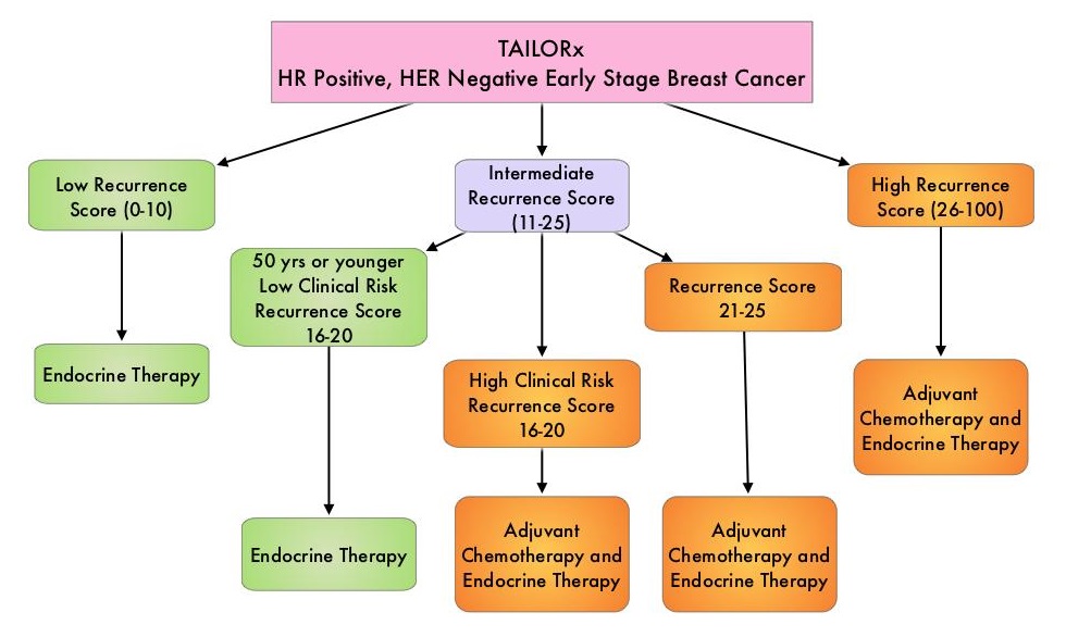
TAILORx ((Trial Assigning Individualized Options for Treatment) is a phase III, randomized, prospective, non-inferiority trial, and is the largest breast cancer treatment trial ever conducted, and the first precision medicine trial ever done, according to the authors. In this study, 10,273 women, 18-75 years of age, with hormone receptor-positive, HER2-negative, axillary node-negative breast cancer were enrolled. Patients had tumors 1.1-5.0 cm in size (or 0.6-1.0 cm and intermediate/high grade). Patients were divided into three groups based on their Recurrence Score. Women with a Low Recurrence Score of 0-10 received endocrine therapy alone and those with a High Recurrence Score of 26-100 received endocrine therapy in combination with standard adjuvant chemotherapy. Patient with Intermediate Recurrence Score of 11-25 (N=6711) were randomly assigned to receive endocrine therapy alone or endocrine therapy and adjuvant chemotherapy. Patients were followed up for 9 years. The Primary endpoint was invasive Disease Free Survival, defined as recurrence of cancer in the breast, regional lymph nodes, and/or distant organs, a second primary cancer in the opposite breast or another organ, or death from any cause. Results reported in June 2018 showed that while most women with an Intermediate Recurrence Score of 11-25 did not benefit from chemotherapy, women 50 years or younger with a Recurrence Score of 16-25 did indeed benefit from adjuvant chemotherapy.

The authors in this publication provided additional results from the same data set showing that adding “Clinical Risk” provides additional prognostic information. The investigators used a binary classification system from the MINDACT trial (Microarray in Node-Negative Disease May Avoid Chemotherapy), which used a 70-gene assay, and divided patients into high or low “Clinical Risk” based on tumor size and histologic grade. Clinical Risk was defined as low if the tumor was 3 cm or less in diameter and had a low histologic grade, 2 cm or less and had an intermediate histologic grade, or 1 cm or less in diameter and had a high grade. The Clinical Risk was defined as high if the low-risk criteria were not met. This additional reporting provided prognostic information about recurrent risk, but not benefits of chemotherapy particularly in the Intermediate Recurrence Score group.TAILORx-HR-Positive-HER-Negative-Early-Stage-Breast-Cancer

Among women who were 50 years of age or younger with a low Recurrence Score, the distant recurrence rate at 9 years was less than 5%, irrespective of Clinical Risk, and about 5% among those with an intermediate Recurrence Score with low Clinical Risk. However in sharp contrast, among women 50 years of age or younger, with high Clinical Risk and an intermediate Recurrence Score who had received endocrine therapy alone, the rate of distant recurrence at 9 years was 12.3%, compared with 6.1% among women who had received adjuvant chemotherapy. It is possible that younger women with a Recurrence Score of 11-25 and high Clinical Risk, receiving endocrine therapy alone, may have been undertreated with Tamoxifen, and the authors speculated that based on previously published studies, adding Ovarian Suppression and an Aromatase Inhibitor might result in risk reduction, equivalent to that observed using adjuvant chemotherapy.

The authors concluded that these new findings complement the original, definitive TAILORx conclusion and integration of genomic (Recurrence Score) and Clinical Risk may provide a more accurate estimate of prognosis for individual patients, than could be provided by either the genomic or clinical information alone. They added that this Clinical Risk stratification facilitates more refined estimates of absolute chemotherapy benefit for women 50 years of age or younger, with a Recurrence Score 16-25. Clinical and Genomic Risk to Guide the Use of Adjuvant Therapy for Breast Cancer. Sparano JA, Gray RJ, Ravdin PM, et al. N Engl J Med 2019;380:2395-2405

BRAFTOVI®, MEKTOVI® and ERBITUX® Triplet Therapy for Patients with BRAF V600E-Mutant Metastatic Colorectal Cancer

SUMMARY: ColoRectal Cancer (CRC) is the third most common cancer diagnosed in both men and women in the United States. The American Cancer Society estimates that approximately 145,600 new cases of CRC will be diagnosed in the United States in 2019 and about 51,020 patients are expected to die of the disease. The lifetime risk of developing CRC is about 1 in 23. Advanced colon cancer is often incurable and standard chemotherapy when combined with anti EGFR (Epidermal Growth Factor Receptor) targeted monoclonal antibodies such as VECTIBIX® (Panitumumab) and ERBITUX® (Cetuximab) as well as anti VEGF agent AVASTIN® (Bevacizumab), have demonstrated improvement in Progression Free Survival (PFS) and Overall Survival (OS). The benefit with anti EGFR agents however is only demonstrable in patients with metastatic CRC (mCRC), whose tumors do not harbor KRAS mutations in codons 12 and 13 of exon 2 (KRAS Wild Type). It is now also clear that even among the KRAS Wild Type patient group about 15-20% have other rare mutations such as NRAS and BRAF mutations, which confer resistance to anti EGFR agents. Patients with stage IV colorectal cancer are now routinely analyzed for extended RAS and BRAF mutations. KRAS mutations are predictive of resistance to EGFR targeted therapy. Approximately 8-15% of all metastatic CRC tumors present with BRAF V600E mutations and BRAF V600E is recognized as a marker of poor prognosis in this patient group. These patients tend to have aggressive disease with a higher rate of peritoneal metastasis and do not respond well to standard treatment intervention. Approximately 20% of the BRAF-mutated population in the metastatic setting has MSI-High tumors, but MSI-High status does not confer protection to this patient group.BRAF-and-MEK-Inhibition-in-MAPK-Pathway

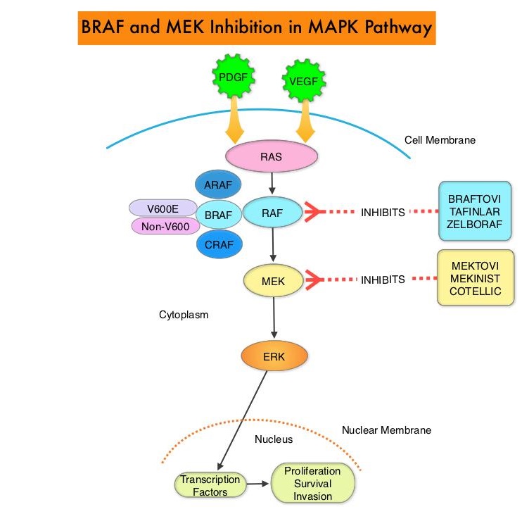
The Mitogen-Activated Protein Kinase pathway (MAPK pathway) is an important signaling pathway which enables the cell to respond to external stimuli. This pathway plays a dual role, regulating cytokine production and participating in cytokine dependent signaling cascade. The MAPK pathway of interest is the RAS-RAF-MEK-ERK pathway. The RAF family of kinases includes ARAF, BRAF and CRAF signaling molecules. BRAF is a very important intermediary of the RAS-RAF-MEK-ERK pathway. The BRAF V600E mutations results in constitutive activation of the MAP kinase pathway. Inhibiting BRAF can transiently reduce MAP kinase signaling. However, this can result in feedback upregulation of EGFR signaling pathway, which can then reactivate the MAP kinase pathway. This aberrant signaling can be blocked by dual inhibition of both BRAF and EGFR. However, BRAF V600E-mutated CRC is inherently less sensitive to BRAF inhibition than melanoma.

The FDA approved BRAFTOVI® (Encorafenib) and MEKTOVI® (Binimetinib) in combination for the first-line treatment of patients with BRAF V600-mutant melanoma, in June 2018. In a recent Phase II study among previously treated patients with BRAF V600E-mutant mCRC, treatment with a combination of BRAFTOVI® plus ERBITUX® resulted in an Objective Response Rate (ORR) of 24%, PFS of 4.2 months, and OS of 9.3 months. These results were significantly better than the standard of care, as well as other BRAF, MEK, and EGFR-inhibitor triplet combinations. Preclinical data suggests that BRAFTOVI® has target binding characteristics that differ from other BRAF inhibitors such as ZELBORAF® (Vemurafenib) and TAFINLAR® (Dabrafenib), with a prolonged target dissociation half-life and higher potency. This may explain the superior efficacy of BRAFTOVI® over other BRAF inhibitors in BRAF V600E-mutated CRC. These encouraging results with the BRAFTOVI® plus ERBITUX® doublet led to the initiation of the Phase III BEACON CRC study.

The BEACON Colorectal Cancer trial is an open-label, randomized, three-arm, Phase III study in which the efficacy and safety of BRAFTOVI® plus ERBITUX® with or without MEKTOVI® was compared with the investigators’ choice of ERBITUX® combined with either Irinotecan or Fluorouracil, Folinic acid, and Irinotecan, in patients with BRAF V600E-mutant mCRC whose disease has progressed after one or two prior regimens. At the time BEACON CRC was initiated, the triplet combination of MEKTOVI®, BRAFTOVI®, and ERBITUX® had not been clinically evaluated. The authors therefore conducted a Safety Lead-In (SLI) to determine the Safety, tolerability, and preliminary efficacy of this triplet combination at the same doses planned for the randomized portion of the trial.

The randomized portion of the trial was ongoing at the time of this analysis. The BEACON trial included patients with mCRC whose tumor tissue was positive for the presence of BRAF V600E mutation. Majority of the patients had right-sided disease as is characteristic of BRAF V600E-mutant mCRC, with a high frequency of nodal and peritoneal metastasis. Liver however, was the most frequent site of metastasis. Enrolled patients must have had progressive disease on one, but no more than two prior treatment regimens, in the metastatic setting. Enrolled patients received BRAFTOVI® 300 mg PO daily plus MEKTOVI® 45 mg PO BID along with ERBITUX® 400 mg/m2 IV, followed by 250 mg/m2 IV weekly every 28 days. The Safety Lead-In was initiated before the randomized portion of the BEACON trial and included 30 patients with disease characteristics and treatment schedule as described above. The Primary end point was Safety, including the incidence of dose-limiting toxicities. Efficacy end points included Overall Response Rate, Progression Free Survival, and Overall Survival.

The median follow up was 18.2 months, and the median time on study drug was 7.9 months. The confirmed Overall Response Rate was 48%, median Duration of Response was 5.5 months, median Progression Free Survival was 8.0 months, and median Overall Survival was 15.3 months. Approximately 79% of responding patients achieved a response within 2 months. The most common adverse events were nausea, diarrhea, fatigue and dermatitis. Approximately 6% of patients experienced serous retinopathy without loss of visual acuity.

It was concluded that in this Safety Lead-In, the combination regimen of BRAFTOVI®, MEKTOVI® and ERBITUX® resulted in promising results compared with available therapies, among patients with previously treated BRAF V600E-mutant mCRC, and if confirmed in the randomized portion of the trial, could become the new standard of care in this patient group. Binimetinib, Encorafenib, and Cetuximab Triplet Therapy for Patients With BRAF V600E–Mutant Metastatic Colorectal Cancer: Safety Lead-In Results From the Phase III BEACON Colorectal Cancer Study. Van Cutsem E, Huijberts S, Grothey A, et al. Journal of Clinical Oncology 2019; 37:1460-1469.

Five-Year Outcomes with TAFINLAR® plus MEKINIST® in Metastatic Melanoma

SUMMARY: It is estimated that in the US, approximately 96,480 new cases of Melanoma will be diagnosed in 2019 and about 7,230 patients are expected to die of the disease. The incidence of Melanoma has been on the rise for the past three decades. Surgical resection with a curative intent is the standard of care for patients with early stage Melanoma, with a 5-year survival rate of 98% for stage I disease and 90% for stage II disease. Patients with locally advanced or metastatic Melanoma historically have had poor outcomes. With the development and availability of immune checkpoint inhibitors and BRAF and MEK inhibitors, this patient group now has significantly improved outcomes. In treatment naïve patients receiving anti-PD-1 therapies such as KEYTRUDA® (Pembrolizumab) or OPDIVO® (Nivolumab) in phase 3 trials, the Progression Free Survival (PFS) rates have ranged from 27-31%, with an Overall Survival (OS) rate of 46% at 4 years. The 5-year OS among patients receiving KEYTRUDA® was 43%, and in those treated with a combination of OPDIVO® plus YERVOY® (Ipilimumab), 4-year PFS and OS rates were 37% and 53%, respectively.BRAF-and-MEK-Inhibition-in-MAPK-Pathway

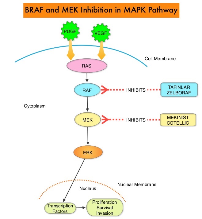
The Mitogen-Activated Protein Kinase pathway (MAPK pathway) is an important signaling pathway which enables the cell to respond to external stimuli. This pathway plays a dual role, regulating cytokine production and participating in cytokine dependent signaling cascade. The MAPK pathway of interest is the RAS-RAF-MEK-ERK pathway. The RAF family of kinases includes ARAF, BRAF and CRAF signaling molecules. BRAF is a very important intermediary of the RAS-RAF-MEK-ERK pathway. BRAF mutations have been detected in 6-8% of all malignancies. The most common BRAF mutation in Melanoma is at the V600E/K site and is detected in approximately 50% of Melanomas, and result in constitutive activation of the MAPK pathway.

TAFINLAR® (Dabrafenib), is a selective oral BRAF inhibitor and MEKINIST® (Trametinib) is a potent and selective inhibitor of MEK gene, which is downstream from RAF in the MAPK pathway. It has been well established that patients who have unresectable or metastatic Melanoma with a BRAFV600E or V600K mutation have prolonged PFS and OS when treated with a combination of BRAF and MEK inhibitors. However, long-term 4 and 5-year clinical outcomes in these patient’s have not been reported.

Two randomized Phase III trials helped address this issue. COMBI-d involved 423 patients randomized to TAFINLAR® plus MEKINIST® (N=211) or to TAFINLAR® plus placebo (N=212). In COMBO-v, 704 patients were randomized to TAFINLAR® plus MEKINIST® (N=352) or to single-agent ZELBORAF® (Vemurafenib; N=352). In a previously published pooled analysis of patients treated in the COMBI-d and COMBI-v trials, 3-year PFS and OS were 23% and 44% respectively. Further, there was a significant association between several baseline factors such as performance status, age, sex, number of organ sites with metastasis, serum LDH level and both PFS as well as OS.

In this review, the researchers analyzed pooled long term survival data from two randomized Phase III COMBI-d and COMBI-v trials, which involved previously untreated, unresectable or metastatic Melanoma patients, with BRAFV600E or V600K mutation, who had received BRAF inhibitor TAFINLAR® 150 mg orally twice daily along with a MEK inhibitor MEKINIST® 2 mg orally once daily. These two trials evaluated the efficacy and safety of TAFINLAR® plus MEKINIST®, as compared with BRAF inhibitor monotherapy. The long term, 5-year survival data from these two trials was reported, along with clinical characteristics of the patients who derived long-term benefit from this treatment. The Primary end points in the COMBI-d and COMBI-v trials were PFS and OS, respectively. The median patient age in these trials was 55 years, 3% of patients had nonmetastatic disease and two-thirds had M1c metastatic disease.

A total of 563 patients (211 in the COMBI-d trial and 352 in the COMBI-v trial) were randomly assigned to receive TAFINLAR® plus MEKINIST®. The PFS rates were 21% at 4 years and 19% at 5 years. The OS rates were 37% at 4 years and 34% at 5 years. The 5-year OS rate was 71% among patients who had a Complete Response and 55% among those who had a normal Lactate Dehydrogenase level plus fewer than three metastatic organ sites at baseline.

It was concluded that first-line treatment with TAFINLAR® plus MEKINIST® led to long-term benefit in approximately one third of the patients who had unresectable or metastatic Melanoma with a BRAF V600E or V600K mutation. The authors added that this is the largest data set and longest follow-up in this patient population treated with BRAF and MEK inhibitors. Five-Year Outcomes with Dabrafenib plus Trametinib in Metastatic Melanoma. Robert C, Grob JJ, Stroyakovskiy D, et al. June 4, 2019. DOI: 10.1056/NEJMoa1904059

FDA Approves First PI3K Inhibitor, PIQRAY®, for Breast Cancer

SUMMARY: The FDA on May 24, 2019 approved PIQRAY® (Alpelisib) in combination with FASLODEX® (Fulvestrant), to treat postmenopausal women and men, with Hormone Receptor (HR) positive, Human Epidermal growth factor Receptor 2 (HER2) negative, PIK3CA-mutated, advanced or metastatic breast cancer (as detected by an FDA-approved test), following progression on or after an endocrine-based regimen. The FDA also approved the companion diagnostic test, therascreen PIK3CA RGQ PCR Kit, to detect the PIK3CA mutation in a tissue and/or a liquid biopsy. Patients who are negative by the therascreen test using the liquid biopsy should undergo tumor biopsy for PIK3CA mutation testing. Breast cancer is the most common cancer among women in the US and about 1 in 8 women (12%) will develop invasive breast cancer during their lifetime. Approximately 268,600 new cases of female breast cancer will be diagnosed in 2019 and about 41,760 women will die of the disease. About 70% of breast tumors express Estrogen Receptors and/or Progesterone Receptors and these patients are often treated with anti-estrogen therapy as first line treatment. However, resistance to hormonal therapy occurs in a majority of the patients.Alpelisib - Mechanism-of-Action

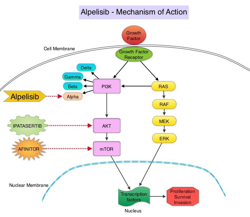
The PhosphoInositide 3-Kinase (PI3K) pathway is an intracellular signaling pathway important in the regulation of cancer cell proliferation and metastasis. PI3K is a lipid kinase and has four distinct isoforms – alpha, beta, gamma and delta, which play a unique role in the survival of different tumor types and establishment of supportive tumor microenvironments. The alpha and beta isoforms are expressed in a wide variety of tissues whereas the gamma and delta isoforms are primarily expressed in hematopoietic cells such as B and T cells. The PI3K alpha isoform is particularly important in breast cancer and plays an important role in tumorigenesis, supporting tumor angiogenesis and stromal interactions, making this a viable target. PIK3CA is an oncogene that codes for the alpha isoform of PI3K, (PI3Kα), more specifically for the alpha isoform of p110. The PI3k pathway is the most frequently altered pathway in human cancers including breast cancer, and has been implicated in disease progression in a significant number of patients with breast cancer. Activation of the PI3K pathway in breast cancer has been associated with resistance to endocrine therapy and disease progression. Approximately 40% of patients with Hormone Receptor positive (HR+), HER2 negative breast cancers, harbor activating mutations in the PIK3CA isoform of PI3K, which is the most common mutation in HR+ breast cancer. Patients with advanced breast cancer harboring PIK3CA mutations typically have a poor prognosis. This provides a strong rationale for targeting the PI3K pathway in breast cancer.

PIQRAY® is an oral, alpha-specific PI3K inhibitor that specifically inhibits PIK3 in the PI3K/AKT kinase signaling pathway. Further, it was shown in preclinical studies that cancer cells with PIK3CA mutations are more sensitive to PIQRAY® than those without the mutation, across a broad range of tumor types. SOLAR-1 clinical trial, which was conducted to test this hypothesis, became the basis for this FDA approval.

SOLAR-1 is a global, double-blind, placebo-controlled, randomized phase III trial, which studied the benefit of PIQRAY® in combination with FASLODEX® (Fulvestrant) among postmenopausal women and men with PIK3CA-mutated HR+/HER2 negative advanced or metastatic breast cancer, who had progressed on or following prior Aromatase Inhibitor (AI) treatment with or without a Cyclin-Dependent Kinase (CDK) 4/6 inhibitor. In this study, 572 patients were randomized in a 1:1 ratio to receive PIQRAY® 300 mg orally daily or placebo once daily, in combination with FASLODEX® 500mg IM on days 1 and 15 of the first cycle and day 1 of each subsequent 28-day cycle. Patients were stratified based on visceral metastases and prior CDK4/6 inhibitor treatment. A total of 341 patients had PIK3CA mutations upon testing of the tumor tissue with 169 patients receiving the PIQRAY® combination and 172 patients receiving FASLODEX® alone. Enrolled patients had received one or more prior lines of hormonal therapy, but no chemotherapy for advanced breast cancer. They had not previously received FASLODEX® or any PI3K, Akt or mTOR inhibitor, and were not on concurrent anticancer therapy. Approximately half of the patients in each treatment group had lung or liver metastases and 6% had received prior CDK4/6 inhibitor therapy. The Primary endpoint was Progression Free Survival (PFS) for patients with the PIK3CA mutation. Secondary endpoints included Overall Survival (OS), Overall Response Rate (ORR), Clinical Benefit Rate, Health-Related Quality of Life, Efficacy in PIK3CA non-mutant cohort, Safety and Tolerability.

The Primary endpoint was met and at a median follow up of 20 months, the PFS was nearly twice as long in patients with PIK3CA mutations randomized to PIQRAY® plus FASLODEX® compared to the placebo plus FASLODEX® group. The median PFS was 11.0 months in the PIQRAY® group compared to 5.7 months in the placebo group (HR=0.65; P=0.00065). In patients with measurable, PIK3CA-mutated advanced breast cancer (N=262), the Overall Response Rate was 36% for the PIQRAY® plus FASLODEX® group versus 16% for placebo plus FASLODEX® group (P=0.0002). There was no significant PFS benefit noted in the PIK3CA-nonmutant patient group receiving PIQRAY® plus FASLODEX® The most frequent toxicities with PIQRAY® were hyperglycemia which could be managed with Metformin, nausea, decreased appetite and skin rash.

It was concluded that PIQRAY® given along with FASLODEX® significantly improved Progression Free Survival compared to Placebo plus FASLODEX® with manageable toxicities. The authors commented that this is the first study to show statistically significant, clinically meaningful PFS improvement with an alpha-specific PI3K inhibitor in PIK3CA-mutated HR+, HER2 negative advanced breast cancer, highlighting the importance of clinical genomics in advanced breast cancer. PIQRAY® is also the first novel drug approved under the Real-Time Oncology Review pilot program. It however remains unclear whether PIQRAY® should be incorporated into the current treatment paradigm upfront, along with endocrine therapy and a CDK 4/6 inhibitor, or sequentially following disease progression on a combination of endocrine therapy and a CDK 4/6 inhibitor. Alpelisib (ALP) + fulvestrant (FUL) for advanced breast cancer (ABC): results of the phase 3 SOLAR-1 trial. André F, Ciruelos EM, Rubovszky G, et al. Presented during the Presidential Symposium 1 at: 2018 ESMO Congress; October 19-23; Munich, Germany. Abstract LBA3_PR.

Liquid Biopsy Accurate, Reliable and Rapid in Identifying Biomarker Mutations in Newly Diagnosed Advanced Lung Cancer

SUMMARY: The American Cancer Society estimates that for 2019 about 228,150 new cases of lung cancer will be diagnosed and 142,670 patients will die of the disease. Non Small Cell Lung Cancer (NSCLC) accounts for approximately 85% of all lung cancers. Patients with newly diagnosed metastatic NSCLC are often tested for guideline-recommended genomic biomarkers which include both predictive biomarker mutations such as EGFR, ALK, ROS1, BRAF, RET, MET, ERBB2, as well as prognostic biomarker mutation such as KRAS.

The application of precision medicine with targeted therapy requires detection of molecular abnormalities in a tissue biopsy specimen. However, if testing is not done with a comprehensive assay, such as Next-Generation Sequencing and is done in successive steps one test after another, tissue sample can be depleted, with not enough tissue left for testing of all biomarkers. Following progression or recurrence, archived biopsy specimens may not be helpful, as it is important to identify additional mutations in the tumor at the time of recurrence or progression, in order to plan appropriate therapy. Further, recurrent tumors may be inaccessible for a safe biopsy procedure or the clinical condition of the patient may not permit a repeat biopsy. Additionally, the biopsy itself may be subject to sampling error due to tumor heterogeneity. Genotyping circulating cell-free tumor DNA (cfDNA) in the plasma can potentially overcome the shortcomings of repeat biopsies and tissue genotyping, allowing the detection of many more targetable gene mutations, thus resulting in better evaluation of the tumor genome landscape.

The Noninvasive versus Invasive Lung Evaluation (NILE) trial is a prospective, multicenter study conducted to demonstrate the noninferiority of comprehensive cell-free DNA (cfDNA) relative to standard-of-care traditional tissue genotyping tests, to identify guideline-recommended genomic biomarkers, in patients with metastatic NSCLC. The authors in this study enrolled 282 newly diagnosed patients at 28 North American centers, with previously untreated, nonsquamous, metastatic NSCLC undergoing standard-of-care tissue genotyping. Enrolled patients submitted a pretreatment blood sample for cfDNA analysis utilizing a CLIA-certified comprehensive 73-gene next generation sequencing panel (Guardant360®). Over 80% of the enrolled patients were white and over 50% were female.

The liquid biopsy utilizing Guardant360®, detected biomarker mutations at a rate similar to standard-of-care tissue genotyping tests, in the enrolled patients, meeting the Primary study objective. At least one of the guideline-recommended genomic biomarkers was detected in 60 patients (21.3%) using tissue-based tests and in 77 patients (27.3%) by cfDNA utilizing Guardant360® (P<0.0001). The detection rate was increased by 48% when Guardant360® was utilized for cfDNA analysis and this included those with negative, not assessed, or Quantity Not Sufficient (QNS) results in tissue. In addition, the Positive Predictive Value was 100% for cfDNA versus tissue genotyping, for FDA approved targets such as EGFR, ALK, ROS1, and BRAF mutations. There are agents already approved by the FDA to treat this patient population. The median turnaround time was significantly lower for cfDNA, compared to tissue genotyping (9 versus 15 days; P <0.0001).

The authors concluded that in this largest cfDNA study among patients with previously untreated advanced NSCLC, cfDNA successfully detected seven biomarker mutations noninvasively, significantly faster than tissue genotype testing, and was also able to rescue biomarker mutation positive patients who had non-diagnostic tissue results. They added that the findings in this study confirms similar findings from Europe and demonstrates the clinical utility of cfDNA in newly diagnosed metastatic NSCLC. Clinical utility of comprehensive cell-free DNA (cfDNA) analysis to identify genomic biomarkers in newly diagnosed metastatic non-small cell lung cancer (mNSCLC). Leighl N, Page RD, Raymond VM, et al. Presented at: AACR Annual Meeting April 2, 2019; Philadelphia, USA.

Anaplastic Lymphoma Kinase Inhibition in Non Small Cell Lung Cancer

SUMMARY:The therapeutic target of interest is an aberrant fusion gene, EML4-ALK. EML4 (echinoderm microtubule-associated protein-like 4) – ALK (anaplastic lymphoma kinase) is a fusion-type oncoprotein and is tyrosine kinase. This oncoprotein/tyrosine kinase is found in 2-7% of all Non Small Cell Lung Cancers (NSCLC) and is generated due to an inversion in the short arm of chromosome 2. This oncoprotein is more prevalent in patients with adenocarcinoma who have little or no exposure to tobacco. Tyrosine kinases normally play an important role in cellular proliferation and differentiation. However with point mutations, translocation/rearrangement and amplifications of their respective genes, these tyrosine kinases can potentially cause malignancy. Such is the case with mutations or translocations of the Anaplastic Lymphoma Kinase gene (ALK). In an article published in the October 28, 2010 issue of the NEJM, Crizotinib an oral small molecule tyrosine kinase inhibitor of ALK tyrosine kinase resulted in an overall response rate of 57% in patients who had progressed on prior therapies. Stable disease was noted in 33% of the patients. This is remarkable considering that the response rates in this patient population treated with second line chemotherapy is around 10-15%. As we move forward, it is very likely that genotyping patients and tailoring therapy accordingly, will become standard practice. N Engl J Med 2010; 363:1693-1703