FDA Approves YONDELIS® for Soft Tissue Sarcomas

SUMMARY: The U.S. FDA approved YONDELIS® (Trabectedin) for the treatment of patients with unresectable or metastatic Liposarcoma or Leiomyosarcoma, who have received a prior Anthracycline-containing regimen. Soft Tissue Sarcomas are a heterogeneous group of tumors of mesenchymal origin with over 50 different histological variants. The American Cancer Society estimates that in 2015, about 11,930 new soft tissue sarcomas will be diagnosed in the United States and 4,870 patients will die of the disease. The most common types of Soft Tissue Sarcomas in adults are undifferentiated pleomorphic sarcoma (previously called Malignant Fibrous Histiocytoma), Liposarcoma, and Leiomyosarcoma. Chemotherapy is the standard of care for advanced Soft Tissue Sarcomas. Following first line therapy with Anthracycline or Ifosfamide based chemotherapy regimens, Response Rates are low and second line treatment options are limited. YONDELIS® (Trabectedin) originally isolated from the Caribbean sea sponge (Ecteinascidia turbinate) is a synthetic alkaloid that binds to the minor groove of DNA and induces apoptosis by damaging the DNA.

Based on promising phase II trials, a randomized, open-label, multicenter, phase III trial was conducted, in which 518 patients with unresectable, locally advanced or metastatic Liposarcoma or Leiomyosarcomas were randomly assigned in a 2:1 to receive either YONDELIS® or Dacarbazine. YONDELIS® was dosed at 1.5 mg/m2, administered as an intravenous infusion over 24 hours (N=345) and Dacarbazine was dosed at 1000 mg/m2 administered as an intravenous infusion over 20 to 120 minutes (N=173) and the treatment was given once every 3 weeks. The median age was 56 years and enrolled patients were heavily pretreated and had received prior Anthracycline containing chemotherapy regimens. Close to 90% of the patients had received at least two prior lines of chemotherapy. Patients were stratified by Soft Tissue Sarcoma subtype (Leiomyosarcoma vs Liposarcoma), ECOG performance status, and number of prior chemotherapy regimens. The primary end point was Overall Survival (OS) and secondary end points included Progression Free Survival (PFS), Time To Progression, Objective Response Rate, Duration of Response, as well as safety.

This study demonstrated a statistically significant improvement in PFS in the YONDELIS® group with a 45% reduction in the risk of disease progression or death compared with Dacarbazine (HR= 0.55; P<0.001). The median PFS was 4.2 and 1.5 months in the YONDELIS® and Dacarbazine groups, respectively. This benefit was noted in patients with both Leiomyosarcoma and Liposarcoma. The greatest benefit in median PFS (5.6 months vs 1.5 months with YONDELIS® vs Dacarbazine, respectively) was noted in the myxoid or round cell Liposarcomas, which are considered as translocation-related sarcomas. This additional benefit can be explained based on the direct inhibition by YONDELIS®, of the chimeric FUS-CHOP translocation-generated oncoprotein, which regulates transcriptional activity in myxoid or round cell Liposarcomas. There was however no significant difference in the median Overall Survival between YONDELIS® and Dacarbazine (12.9 months vs 12.4 months, P=0.37). This may be due to crossover of close to 30% of patients who progressed on Dacarbazine to Receptor Tyrosine Kinase Inhibitor, VOTRIENT® (Pazopanib), which was approved in the U.S. during the course of this study. Most of patients who benefited from YONDELIS®, experienced stable disease as their best response for longer durations, compared to those with Dacarbazine (51% vs 35%).

The most common adverse reactions (20% or more) with YONDELIS® were nausea, fatigue, vomiting, constipation, decreased appetite, diarrhea, peripheral edema, dyspnea, and headache. The most common grade 3 to 4 adverse effects were myelosuppression and transient elevation of transaminases. The authors concluded that YONDELIS® significantly improves Progression Free Survival with superior disease control compared to Dacarbazine, in patients with heavily pretreated, advanced Liposarcoma and Leiomyosarcoma. Efficacy and Safety of Trabectedin or Dacarbazine for Metastatic Liposarcoma or Leiomyosarcoma After Failure of Conventional Chemotherapy: Results of a Phase III Randomized Multicenter Clinical Trial. Demetri GD, von Mehren M, Jones RL, et al. J Clin Oncol, doi: 10.1200/JCO.2015.62.4734.

IMLYGIC® a Novel Oncolytic Immunotherapy Demonstrates Significant Responses in Advanced Melanoma

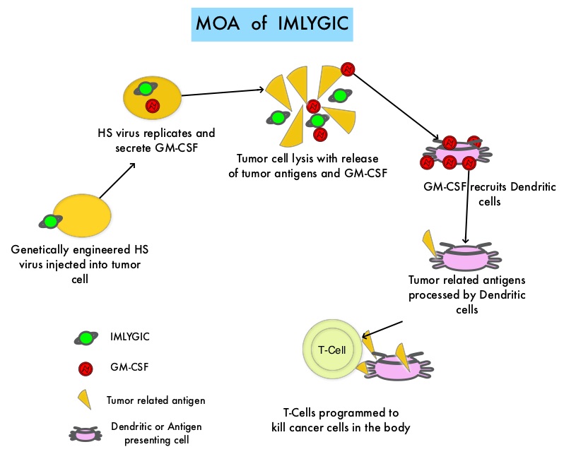
SUMMARY: The FDA on October 27, 2015 approved IMLYGIC® (Talimogene laherparepvec or T-VEC), the first FDA-approved oncolytic virus therapy, for the treatment of melanoma lesions in the skin and lymph nodes. The American Cancer Society’s estimates that for 2015, approximately 74,000 new melanomas will be diagnosed in the United States and about 10,000 people are expected to die of the disease. IMLYGIC® is a genetically modified, herpes simplex virus type 1–derived oncolytic immunotherapy designed to induce both local and systemic immune responses. Following injection directly into melanoma lesions, IMLYGIC® selectively replicates within tumors and produces an immunostimulatory protein called Granulocyte-Macrophage Colony Stimulating Factor (GM-CSF). IMLYGIC® causes cell lysis resulting in the release of tumor-derived antigens, which along with the local GM-CSF, recruit and activate antigen-presenting cells, with subsequent induction of tumor-specific T-cell responses. The enhanced systemic antitumor immune response against tumor-derived antigens, eradicates tumor cells elsewhere in the body.

Previously reported single arm phase II study with IMLYGIC® demonstrated an Overall Response Rate (ORR) of 26% in patients with stage IIIC to IV melanoma, with responses noted in both injected and un-injected lesions, including visceral lesions. Oncovex (GM-CSF) Pivotal Trial in Melanoma (OPTiM) is a phase III study in which 436 patients with stage IIIB to IV melanoma, with injectable melanoma lesions that could not be surgically resected, were randomly assigned in a 2:1 ratio to receive intralesional IMLYGIC® (N=295) or subcutaneous GM-CSF (N=141). The enrolled patient’s melanoma lesions in the skin and lymph nodes were treated with IMLYGIC® or a comparator therapy and Injection into visceral lesions was not allowed. Patients in the IMLYGIC® group received a series of injections into the melanoma lesions. Following the initial injection, a second dose was administered three weeks later, followed by additional doses every two weeks for at least six months, unless other treatment was required or until there are were no remaining injectable lesions to treat. Patients in the GM-CSF group received 125 micrograms/m2 subcutaneously once daily for 14 days in 28-day cycles. The median age of patients in the study was 63 years. The primary end point was Durable Response Rate (DRR- Objective Response lasting continuously for 6 months or more). Secondary end points included Overall Survival (OS) and Overall Response Rate.

The DRR was significantly higher among patients receiving IMLYGIC® than among those given GM-CSF (16.3% vs 2.1%; P<0.001). The Overall Response Rate was also higher with IMLYGIC® compared to GM-CSF (26.4% vs 5.7%; P<0.001). Approximately 11% of patients receiving IMLYGIC® experienced a Complete Response compared to less than 1% for those receiving GM-CSF. The median Time to Treatment Failure was 8.2 months with IMLYGIC® and 2.9 months with GM-CSF (HR=0.42) and median Overall Survival was 23.3 months and 18.9 months, respectively (HR=0.79; P=0.051). In an exploratory subset analysis, the benefit with IMLYGIC® was more pronounced among patients with stage IIIB, IIIC, or IV M1a disease, as well as among patients who were treatment naïve. The most common side effects were fatigue, chills, fever, nausea, flu-like symptoms and pain at the injection site. IMLYGIC® should not be given to individuals with suppressed immune systems or who are pregnant because IMLYGIC® is a modified live oncolytic herpes virus therapy and herpes virus infection can potentially occur.

The authors concluded that IMLYGIC® is the first oncolytic immunotherapy to demonstrate therapeutic benefit in patients with advanced unresectable melanoma and may be another novel therapeutic option for patients with metastatic melanoma. Combining IMLYGIC® with T-Cell checkpoint inhibitor, YERVOY® (Ipilimumab) is presently being explored and thus far has shown encouraging results with minimal added toxicities. Talimogene Laherparepvec Improves Durable Response Rate in Patients With Advanced Melanoma. Andtbacka RH, Kaufman HL, Collichio F, et al. Published online before print May 26, 2015, doi: 10.1200/JCO.2014.58.3377

ONIVYDE® in Combination with 5-FU and Leucovorin Improves Overall Survival in Metastatic Pancreatic Carcinoma

SUMMARY: The FDA on October 22, 2015 approved ONIVYDE® (Irinotecan liposome injection) administered in combination with Fluorouracil (5-FU) and Leucovorin , for the treatment of patients with metastatic adenocarcinoma of the Pancreas, whose disease has progressed following GEMZAR® (Gemcitabine) based therapy. The American Cancer Society estimates that in 2015, close to 49,000 people will be diagnosed with pancreatic cancer in the United States and over 40,000 people will die of the disease. Some important risk factors for Pancreatic cancer include increasing age, obesity, smoking history, genetic predisposition, exposure to certain dyes and chemicals, heavy alcohol use and pancreatitis. The best chance for long term survival is complete surgical resection, although this may not be feasible in a majority of the patients, as they present with advanced disease at the time of diagnosis. Based on the National Cancer Data Base, the 5 year observed survival rate for patients diagnosed with exocrine cancer of the Pancreas is 14% for those with Stage IA disease and 1% for those with Stage IV disease.

ONIVYDE® is a novel nanoliposomal encapsulation of Irinotecan, a topoisomerase 1 inhibitor. It is designed to optimize the delivery of Irinotecan, by extending the duration of circulation of the drug in the body and preferentially activating the drug within the tumor tissues, to achieve higher levels of the active cytotoxic drug metabolite, SN-38. This approach reduces the toxicity of Irinotecan to normal tissues while maintaining or increasing its anti-tumor efficacy.

NAPOLI-1 is an open-label phase III study in which 417 patients with Gemcitabine-refractory metastatic Pancreatic adenocarcinoma were randomly assigned in a 1:1:1 ratio to receive either ONIVYDE® monotherapy, ONIVYDE® plus 5-FluoroUracil (5-FU) and Leucovorin or 5-FU with Leucovorin (control group). Sixty one percent (61%) of patients had cancer in the head of the Pancreas and 68% had liver metastases. Treatment consisted of ONIVYDE® 120 mg/m2 IV over 90 minutes every 3 weeks in Group A, ONIVYDE® 80 mg/m2 IV given over 90 minutes followed by 5-FU 2400 mg/m2 given over 46 hours and racemic Leucovorin 400 mg/m2 IV given over 30 minutes every 2 weeks in Group B and 5-FU 2000 mg/m2 IV given over 24 hours plus racemic Leucovorin 200 mg/m2 IV given over 30 minutes weekly for 4 weeks followed by 2 weeks of rest in Group C (Control group).

Each of the two ONIVYDE® containing groups was compared with the 5FU/Leucovorin control group. The primary study endpoint was Overall Survival and secondary endpoints included Progression Free Survival (PFS) and Overall Response Rate (ORR). The combination of ONIVYDE®, 5-FU and Leucovorin resulted in a median OS of 6.1 months compared with 4.2 months with 5-FU and Leucovorin alone (HR = 0.67; P=0.012). The median PFS was 3.1 months for the ONIVYDE® combination compared with 1.5 months with 5-FU and Leucovorin alone (HR= 0.55; P=0.0001). The ORR was low in both treatment groups (7.7% vs 0.8%). ONIVYDE® montherapy was not superior, compared with 5-FU and Leucovorin and was associated with more side effects compared to the combination regimen. In an expanded, pre-specified analyses, patients who received at least 80% of the target dose in the first 6 weeks experienced an even greater Overall Survival benefit (43% improvement) with ONIVYDE® combination, compared with 5-FU and Leucovorin alone (8.9 months vs 5.1 months, HR=0.57, P=0.011).

The most common grade 3/4 adverse events with ONIVYDE® plus 5-FU and Leucovorin were neutropenia, fatigue, diarrhea and vomiting. The authors concluded that a combination of ONIVYDE® plus 5-FU and Leucovorin significantly improved Overall Survival compared to 5-FU and Leucovorin with manageable toxicities. Expanded analyses of Napoli-1: Phase 3 study of MM-398 (nal-IRI), with or without 5-fluorouracil and leucovorin, versus 5-fluorouracil and leucovorin, in metastatic pancreatic cancer (mPAC) previously treated with gemcitabine-based therapy. Chen L, Von Hoff DD, Li C, et al. J Clin Oncol 33, 2015 (suppl 3; abstr 234)

Targeting CD38 with Daratumumab Monotherapy in Multiple Myeloma

SUMMARY: The FDA recently assigned a priority review designation to Daratumumab, as a treatment for patients with double refractory Myeloma. Multiple Myeloma is a clonal disorder of plasma cells in the bone marrow and the American Cancer Society estimates that in the United States, close to 27,000 new cases will be diagnosed in 2015 and 11,240 will die of the disease. The choice of first line therapy for patients with Myeloma may depend on transplant eligibility, comorbid conditions, cytogenetics and patient performance status. Following progression on first line therapy, single agent therapy with KYPROLIS® (Carfilzomib) has an Objective Response Rate of 24%, with a median duration of response of 7.4 months, in patient groups refractory to both VELCADE® (Bortezomib) and REVLIMID® (Lenalidomide). POMALYST® (Pomalidomide) in combination with Dexamethasone is superior to high dose Dexamethasone alone, with a significant improvement in the Progression Free Survival and Overall Survival, in patients with relapsed and refractory Multiple Myeloma. More recently, data became available for three triplet regimens in patients with relapsed Myeloma – 1) The addition of KYPROLIS® to REVLIMID® and Dexamethasone resulted in significant improvement in Progression Free Survival, when compared with REVLIMID® and Dexamethasone alone, in patients with relapsed Multiple Myeloma 2) A combination of FARYDAK® (Panobinostat), a histone deacetylase (HDAC) inhibitor, VELCADE® and Dexamethasone significantly improves Progression Free Survival in patients with relapsed and refractory Multiple Myeloma when compared to VELCADE® and Dexamethasone 3) Elotuzumab (HuLuc63), a monoclonal antibody that binds to the Signal Lymphocyte Activation Molecule – SLAMF7 protein (CS1, CD319), when added to REVLIMID® and Dexamethasone, reduced the risk of disease progression by 30% in patients with Relapsed/Refractory Multiple Myeloma, when compared with REVLIMID® and Dexamethasone.

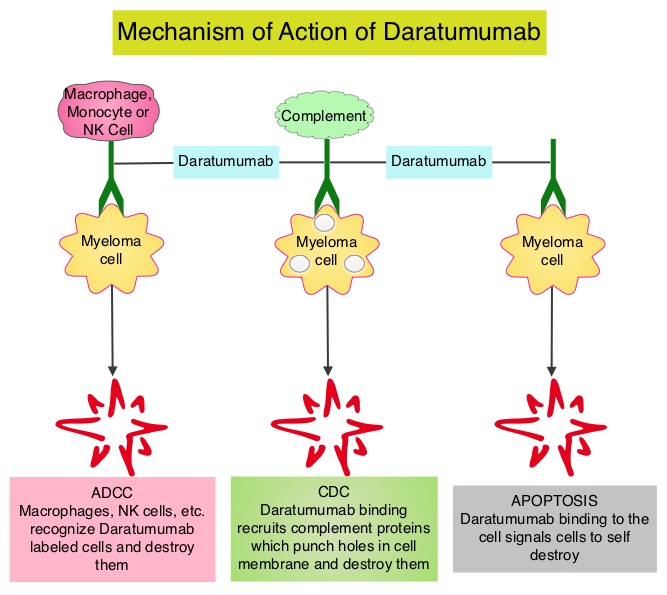
Daratumumab is a human IgG1 antibody that targets CD38, a transmembrane glycoprotein abundantly expressed on malignant plasma cells and with low levels of expression on normal lymphoid and myeloid cells. Daratumumab exerts its cytotoxic effect on myeloma cells by multiple mechanisms, including Antibody Dependent Cellular Cytotoxicity (ADCC), complement mediated cytotoxicity and direct apoptosis. Based on the anti-myeloma activity of Daratumumab in preclinical studies, a phase 1–2 clinical trial was conducted which involved patients with relapsed and refractory Myeloma, who were refractory to two or more prior lines of therapy. Patients in this study had received a median of four previous lines of therapy, 79% of the patients had disease that was refractory to their most recent therapy, including proteasome inhibitors and immunomodulators and 76% of the patients had also undergone autologous stem cell transplantation. This trial included a dose-expansion phase in which 30 patients received Daratumumab 8 mg/kg and 42 patients received 16 mg/kg, given once weekly for 8 doses, twice monthly for 8 doses, and monthly for up to 24 months. The primary end point was safety determined by frequency and severity of adverse events. Secondary end points included Objective Response Rate, duration of response, time to disease progression, Progression Free Survival, Overall Survival and pharmacokinetics.

It was noted that in the cohort that received Daratumumab 16 mg/kg, the overall response rate was 36% with a median Progression Free Survival of 5.6 months and 65% of the patients who had a response did not have disease progression at 12 months.

Infusion related reactions were mild and only 1% had grade 3 events. The other most common adverse events were pneumonia and thrombocytopenia. The authors concluded that Daratumumab has significant single agent activity in a very heavily pretreated and refractory Myeloma patient population, with a favorable safety profile. It remains to be seen if Daratumumab will be an important component of the induction, consolidation and maintenance phases, in the Myeloma treatment landscape. Targeting CD38 with Daratumumab Monotherapy in Multiple Myeloma. Lokhorst HM, Plesner T, Laubach JP, et al. N Engl J Med 2015; 373:1207-1219

Molecular Markers Can Predict Response to Neoadjuvant Cisplatin-based Chemotherapy in Muscle Invasive Bladder Cancer

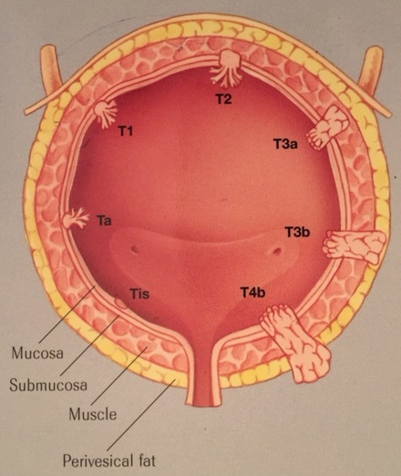
SUMMARY: The American Cancer Society estimates that in the United States for 2015, about 74,000 new cases of bladder cancer will be diagnosed and approximately 16,000 patients will die of the disease. A third of the patients initially present with locally invasive or metastatic disease. Even though radical cystectomy was considered the standard of care for patients with localized Muscle Invasive Bladder Cancer (MIBC), two large randomized trials and two meta-analysis have shown greater survival benefit with neoadjuvant Cisplatin-based chemotherapy combinations for patients with MIBC, compared to surgery alone. However, not all patients with MIBC benefit from neoadjuvant Cisplatin based therapy, with only 25-50% attaining a pathologic response. The authors in this publication evaluated the role of genetic testing and genetic biomarkers prior to chemotherapy, in order to select the appropriate group of patients who would benefit from neoadjuvant Cisplatin based chemotherapy and thereby exclude those who are unlikely to benefit from chemotherapy.

Muscle Invasive Bladder Cancer (MIBC) patients from two prospective multicenter clinical trials of Cisplatin-based neoadjuvant chemotherapy were included and pretreatment MIBC samples were prospectively collected and this provided the discovery and validation sets. DNA from pre-treatment tumor tissue was sequenced for all coding exons of 287 cancer related genes and was analyzed for presence of base substitutions, indels, copy number alterations and selected rearrangements. The discovery set allowed for improved prognostic strength, by combining existing known high risk genetic markers with other novel risk markers. The validation set validated these markers. This study included data from 34 patients in the discovery cohort and 24 patients in the validation cohort.

It was noted that patients with a pathologic Complete Response had more alterations in the genes compared to those with residual tumor, both in the discovery (P= 0.024) and validation (P=0.018) sets. Further, in the discovery set, alteration in one or more of the three DNA repair genes ATM, RB1, and FANCC predicted pathologic Complete Response (P<0.001; 87% sensitivity, 100% specificity) as well as improved Overall Survival (P=0.007). DNA repair is one of the mechanisms postulated to contribute to Cisplatin chemo-resistance. This test remained predictive for pathologic Complete Response in the validation set (P=0.033), with a trend towards improved Overall Survival (P=0.055).

The authors concluded that defective DNA repair renders tumors sensitive to Cisplatin and genomic alterations in the DNA repair-associated genes ATM, RB1, and FANCC predict response and clinical benefit for MIBC, treated with Cisplatin-based chemotherapy. Defects in DNA Repair Genes Predict Response to Neoadjuvant Cisplatin-based Chemotherapy in Muscle-invasive Bladder Cancer. Plimacka ER, Dunbracka RL, Brennan TA, et al. European Urology 2015;68:959-967

YERVOY® and OPDIVO® Combination Superior to YERVOY® Monotherapy in Advanced Melanoma

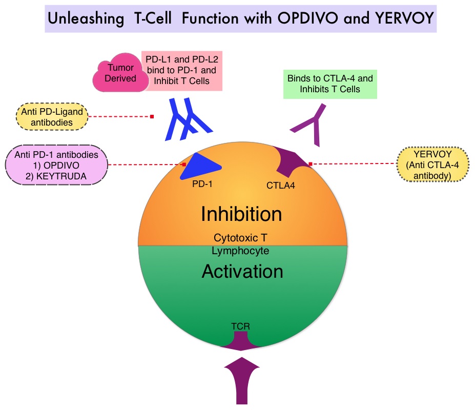
SUMMARY: The FDA on September 30, 2015 granted accelerated approval to OPDIVO® (Nivolumab) in combination with YERVOY® (Ipilimumab), for the treatment of patients with BRAF V600 wild-type, unresectable or metastatic melanoma. The American Cancer Society’s estimates that for 2015, approximately 74,000 new melanomas will be diagnosed in the United States and about 10,000 people are expected to die of the disease. The understanding of the Immune checkpoints has lead to the development of novel immunotherapies. Immune checkpoints or gate keepers are cell surface inhibitory proteins/receptors that are expressed on activated T cells. They harness the immune system and prevent uncontrolled immune reactions. Survival of cancer cells may be related to their ability to escape immune surveillance, by inhibiting T lymphocyte activation. With the recognition of Immune checkpoint proteins and their role in suppressing antitumor immunity, antibodies have been developed that target the membrane bound inhibitory Immune checkpoint proteins/receptors such as CTLA-4 (Cytotoxic T-Lymphocyte Antigen 4), PD-1(Programmed cell Death-1), etc. By doing so, the T cells are unleashed, resulting in T cell proliferation, activation and a therapeutic response. YERVOY® is a fully human immunoglobulin G1 monoclonal antibody, that blocks Immune checkpoint protein/receptor CTLA-4 and has been shown to prolong Overall Survival in patients with previously treated, unresectable or metastatic melanoma. OPDIVO® is a fully human, immunoglobulin G4 monoclonal antibody that binds to the PD-1 receptor and blocks its interaction with PD-L1 and PD-L2, thereby undoing PD-1 pathway-mediated inhibition of the immune response. OPDIVO® significantly improves Objective Response Rates, in patients with advanced melanoma. Previously published phase I studies demonstrated high Objective Response Rates, including Complete Responses, among patients with advanced melanoma, when treated with a combination of YERVOY® and OPDIVO®.

In this double-blind phase II trial, 142 treatment naïve patients with metastatic melanoma were randomly assigned in a 2:1 ratio to receive either OPDIVO® in combination with YERVOY® (N=95) or YERVOY® plus placebo (N=47). Patients in the OPDIVO® plus YERVOY® group received OPDIVO® 1 mg/kg and YERVOY® 3 mg/kg IV every 3 weeks for four doses followed by OPDIVO® 3 mg/kg every 2 weeks until disease progression or unacceptable toxicity. Patients in the YERVOY® and placebo group received YERVOY® 3 mg/kg and placebo IV every 3 weeks for four doses followed by placebo. At the time of disease progression, patients in the YERVOY® group were offered OPDIVO® 3 mg/kg every 2 weeks. Patients were stratified according to BRAF mutation status (wild type versus mutation-positive). The median age was 65 years and majority of patients were males. The primary end point was Objective Response Rate among patients with BRAF V600 wild type tumors. The authors noted that in BRAF wild type tumors, the overall Response Rate was 61% with the combination treatment versus 11% for YERVOY® alone (P<0.001). The Complete Response rate was 22% for the combination therapy vs 0% for YERVOY®. The median duration of response was not reached in either treatment groups, with an ongoing response rate of 82% in the combination group and 75% in the YERVOY® plus placebo group. The median Progression Free Survival was not reached with the combination therapy and was 4.4 months in the YERVOY® plus placebo group (HR=0.40; P<0.001). Among patients with BRAF mutation-positive tumors, the overall Response Rate was 52% for the combination therapy group and Complete Response rate was 22%, with similar efficacy compared with the BRAF wild type tumor group. The median Progression Free Survival was 8.5 months for the combination therapy group and 2.7 months for YERVOY® plus placebo group.

Grade 3 or 4 adverse events were reported in 54% of the patients who received the combination therapy as compared with 24% of the patients who received YERVOY® monotherapy and the most common grade 3 or 4 toxicities in the combination therapy group were colitis, diarrhea and elevated transaminases, which resolved with steroids. The authors concluded that a combination of YERVOY® and OPDIVO® is superior to YERVOY® monotherapy, among treatment naïve patients with metastatic melanoma, with significantly greater objective response rate, as well as Progression Free Survival. Nivolumab and Ipilimumab versus Ipilimumab in Untreated Melanoma. Postow MA, Chesney J, Pavlick AC, et al. N Engl J Med 2015; 372:2006-2017

FDA Approves LONSURF®, A Novel Oral Agent for Refractory Colorectal Cancer

SUMMARY: The FDA on September 22, 2015 approved LONSURF® (Trifluridine/Tipiracil) for the treatment of patients with metastatic ColoRectal Cancer (CRC), who have been previously treated with Fluoropyrimidine, Oxaliplatin and Irinotecan-based chemotherapy, an anti-VEGF biological therapy and if RAS wild-type, an anti-EGFR therapy. The American Cancer Society estimates that approximately 133,000 new cases of ColoRectal Cancer (CRC) will be diagnosed in the United States in 2015 and close to 50,000 are expected to die of the disease. Approximately 15-25% of the patients with CRC present with metastatic disease at the time of diagnosis (synchronous metastases) and 50-60% of the patients with CRC will develop metastatic disease during the course of their illness. Patients with metastatic CRC, whose disease has progressed after treatment with standard therapies, have limited therapeutic options available, to treat their disease.

LONSURF® is a combination of two agents – a novel oral nucleoside, Trifluridine and a thymidine phosphorylase inhibitor, Tipiracil hydrochloride. This combination has a unique mechanism of action. Trifluridine, the active ingredient of LONSURF® incorporates into DNA resulting in DNA damage. Degradation of Trifluridine which occurs when taken orally is prevented by Tipiracil hydrochloride.

The RECOURSE study is a pivotal, global, phase III trial, in which 800 patients with metastatic ColoRectal Cancer, refractory to all standard therapies were randomly assigned in a 2:1 ratio to receive either LONSURF® (N=534) or placebo (N=266). Patients received LONSURF® 35mg/m2 or matching placebo orally, twice daily after meals, on Days 1-5 and 8-12 of each 28 day cycle. Treatment was continued until disease progression or unacceptable toxicity. Eligible patients had metastatic ColoRectal Cancer (mCRC), previously treated with chemotherapy and biological therapy, which included Fluoropyrimidine, Oxaliplatin and Irinotecan-based chemotherapy, an anti-VEGF biological therapy and if RAS wild-type, an anti-EGFR therapy. The primary endpoint of this study was overall survival and the secondary endpoint was progression-free survival.

It was noted that LONSURF® significantly improved Overall Survival compared to placebo (7.1 months vs 5.3 months; HR=0.68; P<0.001), with a 32% reduction in the risk of death. LONSURF® also significantly improved Progression Free Survival compared to placebo (HR = 0.47; P<0.001). The most common grade 3 or more adverse events were leukopenia (21%), anemia (18%) and febrile neutropenia (4%), noted in patients receiving LONSURF®. The authors concluded that LONSURF® significantly improved Overall Survival in patients with refractory metastatic ColoRectal Cancer, providing a novel oral therapeutic option for this patient group. Randomized Trial of TAS-102 for Refractory Metastatic Colorectal Cancer. Mayer RJ, Van Cutsem E, Falcone A, et al. N Engl J Med 2015; 372:1909-1919

Preserving Fertility with ZOLADEX® in Breast Cancer

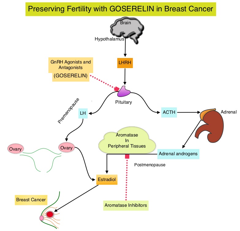
SUMMARY: Breast cancer is the most common cancer among women in the US and about 1 in 8 women (12%) will develop invasive breast cancer during their lifetime. Approximately, 231,840 new cases of invasive breast cancer will be diagnosed in 2015 and over 40,000 women will die of the disease. Approximately 75% of patients with breast cancer are hormone receptor positive (Estrogen Receptor/Progesterone Receptor positive) and this is a predictor of response to endocrine therapy. In premenopausal woman, the ovary is the main source of estrogen production, whereas in postmenopausal women, the primary source of estrogen is the Aromatase enzyme mediated conversion of androstenedione and testosterone to estrone and estradiol in extragonadal/peripheral tissues. Premature Ovarian Failure (POF) is a common unintended consequence of chemotherapy in premenopausal women. Besides of loss of fertility, which can influence treatment decisions in young women, ovarian failure can lead to menopausal symptoms, sexual dysfunction and loss of bone density.

POEMS (Prevention of Early Menopause Study) is a randomized phase III trial designed to evaluate whether the addition of LHRH (Luteinizing Hormone-Releasing Hormone) analog Goserelin (ZOLADEX®), which suppresses the production of estrogens, to Cyclophosphamide based chemotherapy, would reduce POF in breast cancer patients, when compared to chemotherapy alone. Premenopausal patients less than 50 years of age, with hormone receptor negative (ER/PR negative ), Stage I-IIIA breast cancer, scheduled to receive chemotherapy, were randomly assigned to receive standard Cyclophosphamide based chemotherapy with or without monthly ZOLADEX® . Patients in the ZOLADEX® group received 3.6 mg SQ starting 1 week prior to the first dose of chemotherapy.

The primary endpoint was ovarian failure at two years (defined as amenorrhea for the prior 6 months AND post-menopausal FSH level). Other endpoints included pregnancy and survival rates. The median age of the patients was 38 years and median follow up was 4.1 years. Of the 218 evaluable patients, 135 premenopausal women were evaluable for the primary end point. POF rates were 22% in the chemotherapy alone group and 8% in the ZOLADEX® group (P=0.04). When the definition of POF was more liberal to include EITHER amenorrhea or elevated FSH but not both, POF rates were 45% in the chemotherapy alone group and 20% in the ZOLADEX® group (P=0.006). Among the 218 evaluable patients, more women in the ZOLADEX® group achieved at least one pregnancy (21%) compared to 11% in the chemotherapy alone group (P=0.03). Secondary outcomes also favored the ZOLADEX® group with a Disease free Survival (DFS) rate of 78% in the chemotherapy alone group compared with 89% in the ZOLADEX® group (P=0.04) and Overall Survival (OS) rate of 82% in the chemotherapy alone group compared with 92% in the ZOLADEX® group (P=0.05).

The authors concluded that the addition of ZOLADEX® to chemotherapy improved fertility prospects with a lower incidence of Premature Ovarian Failure and more pregnancies. Further, the improved Disease Free Survival and Overall Survival are important additional perks and prevention of Premature Ovarian Failure with ZOLADEX® may be a consideration not only in premenopausal breast cancer patients but also in other malignancies such as lymphomas, when treated with similar chemotherapeutic agents. Goserelin for Ovarian Protection during Adjuvant Chemotherapy for Breast Cancer. Moore HC, Unger JM, Phillips K, et al. N Engl J Med 2015; 372:923-932

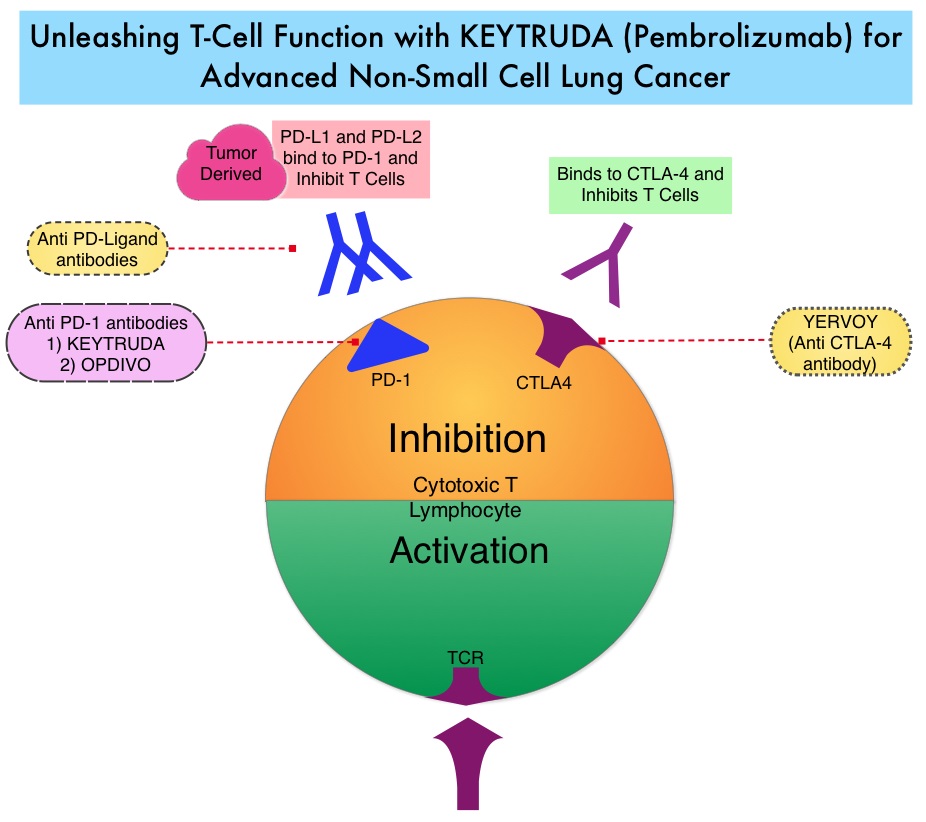
FDA Approves KEYTRUDA® for Advanced Lung Cancer

SUMMARY: The FDA granted accelerated approval to KEYTRUDA® (Pembrolizumab), for the treatment of patients with metastatic Non Small Cell Lung Cancer (NSCLC), whose tumors express Programmed Death Ligand 1 (PD-L1), as determined by an FDA-approved test, following disease progression on or after platinum-containing chemotherapy. Lung cancer is the second most common cancer in both men and women and accounts for about 13% of all new cancers and 27% of all cancer deaths. It is the leading cause of cancer death among both men and women. The American Cancer Society estimates that over 221,200 new cases of lung cancer will be diagnosed in the United States in 2015 and over 158,000 patients will die of the disease. Non Small Cell Lung Cancer (NSCLC) accounts for approximately 85% of all lung cancers.

The treatment paradigm for solid tumors has been rapidly evolving with a better understanding of the Immune checkpoints. Immune checkpoints are cell surface inhibitory proteins/receptors that are expressed on activated T cells. They harness the immune system and prevent uncontrolled immune reactions. Survival of cancer cells in the human body may be to a significant extent, related to their ability to escape immune surveillance, by inhibiting T lymphocyte activation. The T cells of the immune system therefore play a very important role in modulating the immune system. Under normal circumstances, Immune checkpoints or gate keepers, switch off the T cells of the immune system and thereby inhibit an intense immune response. With the recognition of Immune checkpoint proteins and their role in suppressing antitumor immunity, antibodies have been developed, that target the membrane bound inhibitory Immune checkpoint proteins/receptors such as CTLA-4 (Cytotoxic T-Lymphocyte Antigen 4), also known as CD152, PD-1(Programmed cell Death-1), etc. Targeting Immune checkpoints unleashes the T cells, resulting in T cell proliferation, activation and a therapeutic response. KEYTRUDA® is a fully humanized, Immunoglobulin G4, anti-PD-1, monoclonal antibody, that binds to the PD-1 receptor and blocks its interaction with ligands PD-L1 and PD-L2, thereby undoing PD-1 pathway-mediated inhibition of the immune response and unleashing the tumor-specific effector T cells.

In this publication, the authors assessed the efficacy and safety of KEYTRUDA® in patients with advanced NSCLC enrolled in the KEYNOTE-001 phase I trial. Four Hundred and Ninety five (N=495) patients were assigned to either a training group (N=182) or a validation group (N=313) and KEYTRUDA® was administered at three dosages: 2 mg/kg IV every 3 weeks, 10 mg/kg IV every 3 weeks, or 10 mg/kg IV every 2 weeks. Patient responses were assessed every 9 weeks.

The Objective Response Rate (ORR) in the entire study population was 19.4%, the median Duration of Response was 12.5 months, the median Progression Free Survival was 3.7 months and the median Overall Survival was 12.0 months. The PD-L1 (Programmed Death-Ligand 1) expression was evaluated in 204 patients in the validation group by ImmunoHisto Chemistry (IHC) and membrane PD-L1 expression of 50% or more, in tumor cells, was selected as the cutoff. It was noted that among patients with PD-L1 expression in at least 50% of tumor cells, the Objective Response Rate was 45.2%, median Progression Free Survival was 6.3 months and median Overall Survival has not been reached. Responses were not as robust in those patients with tumors demonstrating less than 50% PD-L1 expression, but in those who did respond, the duration of responses were comparable to those with 50% or more PD-L1 expression. KEYTRUDA® was well tolerated overall and the common immune mediated adverse events were infusion reactions, hypothyroidism and pneumonitis.

The authors concluded that KEYTRUDA® showed significant antitumor activity in patients with advanced Non Small Cell Lung Cancer, whose tumor PD-L1expression was 50% or more. Further, the median duration of response exceeded 12 months among responders, regardless of the degree of PD-L1 expression. This study validated that PD-L1 expression in tumors is clearly a marker of response to KEYTRUDA®. Pembrolizumab for the Treatment of Non–Small-Cell Lung Cancer. Garon EB, Rizvi NA, Hui R, et al. N Engl J Med 2015; 372:2018-2028

Breast Cancer Screening Imaging Modalities – A Primer

SUMMARY: Breast cancer is the most common cancer among women in the US and about 1 in 8 women (12%) will develop invasive breast cancer during their lifetime. Approximately, 231,840 new cases of invasive breast cancer will be diagnosed in 2015 and over 40,000 women will die of the disease. Screening mammography complemented by breast self exam and clinical breast exam has resulted in early detection of breast cancer and successful outcomes. Even though mammography is a sensitive screening test, a small percentage of breast cancers may not show up on mammograms but may be palpable on examination by the patient or the clinician. Further, mammograms are less likely to find breast tumors in younger women with dense breast tissue. The following is a brief overview of the imaging techniques for breast cancer screening

Mammography

Mammography is performed by compressing the breast firmly between two plates and utilizes ionizing radiation to image breast tissue. During routine screening, breast tissue from the nipple to the pectoral muscle in the mediolateral oblique and craniocaudal views, is included. The radiation exposure is 4 to 24 mSv per standard two view screening examination. Two view screening is associated with a lower recall rate and lower interval cancer rates than are single-view exams. Breast Imaging Reporting and Data System (BI-RADS) categories are used for reporting mammographic results as follows:

0: Incomplete—needs additional image evaluation and/or prior mammograms for comparison.

1: Negative.

2: Benign.

3: Probably benign.

4: Suspicious.

5: Highly suggestive of malignancy.

6: Known biopsy—proven malignancy.

A digital mammogram is more expensive than screen-film mammography (SFM) and the data can be stored and shared. Compared with film mammography, sensitivity is higher for digital mammography, particularly in women younger than 50 years, but the specificity is either the same or lower than film mammography.

Computer-Aided Detection (CAD) systems increase detection of ductal carcinoma in situ (DCIS) by highlighting suspicious regions in the breast such as clustered microcalcifications and masses in mammograms. There is however no improvement in invasive cancer detection rate and there is an increase in recall rate.

Tomosynthesis

Tomosynthesis, or 3-Dimensional (3-D) mammography involves multiple short-exposure x-rays, from different angles and a three dimensional image is created for better visualization. A combination of 2-D and 3-D mammography has been reported to be more accurate than 2-D mammography alone, with the caveat that the radiation exposure to the patient is essentially doubled. Tomosynthesis in the diagnostic setting is at least as effective as spot compression views, for workup of non-calcified abnormalities, including asymmetries and distortions and may decrease the need for ultrasound testing.

Ultrasonography

Primarily utilized for the diagnostic evaluation of palpable or mammographically detected masses and distinguish solid tumors from cysts. It is a helpful adjunct modality in women with dense breast tissue. Images are created using high frequency sound waves with no radiation exposure. Evidence is lacking to support the use of ultrasound instead of mammography, at any age, in population based breast cancer screening.

Thermography

Thermography uses infrared imaging techniques and identifies temperature changes in the skin as an indicator of an underlying tumor. These changes are displayed in color patterns. The impact of thermography on breast cancer detection or mortality, has not been evaluated in randomized clinical trials and there appears to be no additional benefit for the use of thermography as an adjunct modality, for breast cancer screening.

Magnetic Resonance Imaging

Magnetic Resonance Imaging (MRI) is more sensitive than mammography although the specificity of a breast MRI is lower, resulting in a higher rate of false-positive findings and potentially unnecessary biopsies. Microcalcifications in the breast can be missed by a breast MRI. The American Cancer Society (ACS) recommends an annual MRI as an adjunct to screening mammogram and clinical breast exam in certain groups with increased risk of breast cancer. They include individuals with deleterious genetic mutations such as BRCA1/2 mutation carriers, a strong family history of breast cancer, or several genetic syndromes such as Li-Fraumeni or Cowden disease. MRI may also be used to evaluate the integrity of silicone breast implants, assess palpable masses following surgery or radiation therapy, detect mammographically and sonographically occult breast cancer in patients with axillary nodal metastasis and preoperative planning for some patients with known breast cancer. Breast MRI is performed preferably between days 7-15 of menstrual cycle for premenopausal women, using a dedicated breast coil, with the ability to perform a biopsy under MRI guidance by experienced radiologists, during the same visit.

Molecular Breast Imaging

Molecular Breast Imaging (MBI) involves the injection of technetium-99m (Tc-99m) sestamibi, a radioactive substance, which then allows tumor visualization with a gamma camera. MBI along with mammography significantly increased the cancer detection rate in women with mammographically dense breasts, compared to mammography alone, in a recent study. This new technology is not yet widely available.

References: 1) American Cancer Society recommendations for early breast cancer detection in women without breast symptoms. http://www.cancer.org/cancer/breastcancer 2) National Cancer Institute: PDQ® Breast Cancer Screening. Bethesda, MD