HERIZON-GEA-01 and the Future of HER2-Targeted Therapy in Gastroesophageal Adenocarcinoma

Written by: Benjamin L. Kitchens, MD
Sponsored by: Jazz Pharmaceuticals

Gastroesophageal adenocarcinoma (GEA) includes cancers of the gastroesophageal junction (GEJ) and stomach.  In 2026, U.S. estimates project 31,510 new cases of gastric cancer and 22,530 new cases of esophageal cancer [1,2].  Gastric and GEJ cancer are often diagnosed at advanced stages due to lack of screening programs in the US; this portends a poor prognosis.  The 5-year relative survival for gastric cancer is 78.1% for localized disease, 39.0% for regional disease, and 8.1% for distant metastatic disease [1].  Similarly, esophageal cancer 5-year relative survival rates are 48.6% for localized disease, 29.1% for regional disease, and 5.3% for distant metastatic disease [2].  The poor prognoses for patients with metastatic GEA underscore the need for continued advances in early detection and innovative therapies to improve outcomes.

Biomarker-driven therapy has transformed the treatment landscape for metastatic GEA.  Approximately 14 to 22% of GEAs are HER2-positive [3-5], and HER2-targeted therapy has become central to treatment.  First line therapy for advanced GEA currently consists of FOLFOX plus trastuzumab, with the addition of pembrolizumab for PD-L1 positive (combined positive score ≥ 1) disease, based on the Keynote-811 trial [6].  This trial showed a median overall survival (OS) of 20 months for pembrolizumab added to trastuzumab and chemotherapy, versus 16.8 months for trastuzumab and chemotherapy alone.  Median progression-free survival (PFS) was 10.0 versus 8.1 months, respectively [6].  HER2-directed therapy remains relevant in the second line setting, with the antibody-drug conjugate trastuzumab deruxtecan (T-Dxd) available.  While these treatments reflect impactful progress, there remains a critical need for advanced HER2-directed therapies that deliver deeper and more durable responses.  Zanidatamab is a dual HER2-targeted bispecific immunoglobulin G1-like antibody that binds to two separate extracellular domains of HER2 (domains 2 and 4) [7].  Zanidatamab has already received accelerated approval for previously treated unresectable or metastatic HER2-positive biliary tract cancer, and is under investigation for the treatment GEA [7].

The ongoing Herizon-GEA-01 study is a phase 3 randomized trial evaluating first-line zanidatamab in combination with chemotherapy, plus or minus anti-PD-1 immunotherapy [8].  Participants were 18 years or older with histologically confirmed unresectable, locally advanced, recurrent or metastatic HER2-positive GEA [8].  HER2 positivity consisted of a HER2 immunohistochemistry (IHC) score of 3+, or IHC2+ with positive in situ hybridization.  Other inclusion criteria were an ECOG performance status of 0 or 1, assessable disease defined by RECIST 1.1 criteria, adequate organ function, and left ventricular ejection fraction at least 50%.  Select exclusion criteria were untreated or symptomatic CNS metastases, prior treatment for locally advanced/metastatic GEA, prior HER2-targeted treatment, and prior immunotherapy including anti-PD-1 and anti-PD-L1 agents.

In Herizon-GEA-01, 914 patients were stratified based on geographic region, HER2 status, and ECOG performance status.  Participants were randomly assigned 1:1:1 to one of three treatment arms [8,9].  Arm A patients received trastuzumab plus physician’s choice of chemotherapy (CAPOX or FP).  Arm B received IV zanidatamab 1800mg (weight <70kg)/2400mg (weight ≥70kg) Q3W plus physician’s choice of chemotherapy.  Arm C received IV zanidatamab plus IV tislelizumab 200mg Q3W plus physician’s choice of chemotherapy.

Patients continued treatment until death, disease progression, or unacceptable toxicity.  Chemotherapy could be discontinued after 6 cycles.  The median age in the treatment arms ranged from 62.5 to 64.  Most patients were male (77.3 to 80.8%), ECOG performance status of 1 (55.9 to 61%), metastatic disease (94 to 97.1%), HER2 IHC 3+ (82.6 to 83.1%) and with a PD-L1 tumor area positivity (TAP) score of 1% or greater (58.6 to 61.9%) [10].  Dual primary endpoints were PFS by blinded independent central review and OS.  Secondary endpoints included objective response rate, duration of response, safety, health-related quality of life, and pharmacokinetics.

Interim analysis of HERIZON-GEA-01 demonstrates deeper and more durable responses in the zanidatamab arms than those seen in the trastuzumab plus chemotherapy control arm [10,11].  The median PFS was 12.4 months in both zanidatamab-treated arms compared to 8.1 months in the control arm.  Median overall survival was 24.4 months in the zanidatamab plus chemotherapy arm, and 26.4 in the zanidatamab plus tislelizumab plus chemo arm, compared to 19.2 months in the control arm.  Objective response rates were 65.7% in the control arm, 69.6% in the zanidatamab plus chemotherapy arm, and 70.7% in the zanidatamab plus tislelizumab plus chemotherapy arm.  Median duration of response was 8.3 months in the control arm, 14.3 months in the zanidatamab plus chemotherapy arm, and 20.7 months in the zanidatamab plus tislelizumab plus chemotherapy arm.

Safety data from HERIZON-GEA-01 interim analysis reveals expected results and a manageable zanidatamab toxicity profile.  Diarrhea was the most common adverse event across all three arms, occurring in 82% and 76% of patients in the zanidatamab-treated arms [10].  Other common adverse events across all groups included nausea, vomiting and decreased appetite [11].  Of note, diarrhea prophylaxis was mandatory for patients treated with zanidatamab.  Therefore, appropriate antidiarrheal prophylaxis in the clinic will be very important.  This trial is ongoing and we anticipate further survival data in the future.

This pivotal data from HERIZON-GEA-01 is anticipated to have a meaningful impact on this patient population.  This is particularly in light of the deeper and more long-lasting responses.  Over time, we will see if this translates into statistically significant overall survival and progression-free survival benefits.

References:

  1. Cancer Stat Facts: Stomach Cancer. National Cancer Institute. Surveillance, Epidemiology, and End Results Program.  https://seer.cancer.gov/statfacts/html/stomach.html.  Accessed May 13, 2026.
  2. Cancer Stat Facts: Esophageal Cancer. National Cancer Institute. Surveillance, Epidemiology, and End Results Program.  https://seer.cancer.gov/statfacts/html/esoph.html.  Accessed May 13, 2026.
  3. Van Cutsem E, Bang YJ, Feng-Yi F, et al. HER2 screening data from ToGA: targeting HER2 in gastric and gastroesophageal junction cancer.  Gastric Cancer.  2015;18(3):476-484.  doi:10.1007/s10120-014-0402-y.
  4. Janjigian YY, Werner D, Pauligk C, et al. Prognosis of metastatic gastric and gastroesophageal junction cancer by HER2 status: a European and USA International collaborative analysis.  Ann Oncol.  2012;23(10):2656-2662.  doi:10.1093/annonc/mds104.
  5. Kim WH, Gomez-Izquierdo L, Vilardell F, et al. HER2 Status in Gastric and Gastroesophageal Junction Cancer: Results of the Large, Multinational HER-EAGLE Study.  Appl Immunohistochem Mol Morphol.  2018;26(4):239-245.  doi:10.1097/PAI.0000000000000423.
  6. Janjigian YY, Kawazoe A, Bai Y, et al. Pembrolizumab in HER2-Positive Gastric Cancer.  N Engl J Med. 2024;391(14):1360-1362.  doi:10.1056/NEJMc2408121.
  7. Ziihera ® (zanidatamab-hrii) | Official Website for US Healthcare Professionals. Jazz Pharmaceuticals.  https://www.ziiherahcp.com/.  Accessed May 13, 2026.
  8. A Study of Zanidatamab in Combination With Chemotherapy With or Without Tislelizumab in Subjects With HER2-positive Unresectable Locally Advanced or Metastatic Gastroesophageal Adenocarcinoma (GEA). gov.  https://clinicaltrials.gov/study/NCT05152147.  Accessed May 13, 2026.
  9. Tabernero J, Shen L, Elimova E, et al. HERIZON-GEA-01: Zanidatamab + chemo ± tislelizumab for 1L treatment of HER2-positive gastroesophageal adenocarcinoma.  Future Oncol.  2022;18(29):3255-3266.  doi:10.2217/fon-2022-0595.
  10. Elimova E, Rha SY, Shitara K, et al. Zanidatamab + chemotherapy (CT) ± tislelizumab for first-line (1L) HER2-positive (HER2+) locally advanced, unresectable, or metastatic gastroesophageal adenocarcinoma (mGEA): Primary analysis from HERIZON-GEA-01.  J Clin Oncol.  2026;44(2 suppl):LBA285.  doi:10.1200/JCO.2026.44.2_suppl.LBA285.
  11. Zanidatamab With or Without Tislelizumab Yields Clinically Meaningful Survival Benefit in HER2-Positive Advanced Gastroesophageal Adenocarcinoma. ASCO Daily News.  https://dailynews.ascopubs.org/do/zanidatamab-without-tislelizumab-yields-clinically-meaningful-survival-benefit-her2.  Accessed May 13, 2026.
CheckMate-9ER primary and exploratory findings: baseline characteristics and treatment outcomes, including depth and durability of response with cabozantinib plus nivolumab

CheckMate-9ER primary and exploratory findings: baseline characteristics and treatment outcomes, including depth and durability of response with cabozantinib plus nivolumab

Written by Dr Manojkumar Bupathi
Sponsored by Exelixis, Inc.

Baseline disease characteristics and tumor biology play a pivotal role in shaping outcomes for patients with advanced renal cell carcinoma (aRCC). As treatment paradigms have evolved toward combination strategies, it has become increasingly important not only to demonstrate efficacy in the overall population, but also to consider the potential for the depth, durability, and consistency of responses across clinically relevant subgroups.1,2

The Phase 3 CheckMate-9ER trial established cabozantinib plus nivolumab as a first-line option for patients with aRCC, demonstrating significant improvements in progression-free survival, overall survival, and objective response rate compared with sunitinib. However, beyond these pivotal results, additional insights from descriptive analyses on long-term outcomes and response characteristics are noteworthy to help inform real-world clinical decision-making.3

In this article, we review the key findings from the CheckMate-9ER trial, including updated 5-year follow-up data, and explore post hoc analyses evaluating baseline characteristics associated with response. In particular, we focus on patients achieving complete responses and examine the depth and durability of response to better contextualize the long-term clinical data observed with cabozantinib plus nivolumab.1,4,5

Overview of CheckMate‐9ER
Cabozantinib is a tyrosine kinase inhibitor, targeting MET, AXL, and VEGFR. Based on the results from the CheckMate-9ER trial, a Phase 3, randomized, open-label, head-to-head trial versus sunitinib, cabozantinib + nivolumab was approved for patients with previously untreated aRCC.3,4

We will begin by reviewing the results from the pivotal trial CheckMate-9ER, which enrolled 651 patients with previously untreated aRCC containing a clear-cell component. Participants were randomized 1:1 to receive cabozantinib 40 mg once daily plus nivolumab 240 mg every 2 weeks, or sunitinib 50 mg once daily administered on a 4 weeks on, 2 weeks off schedule.3,4

  • Endpoints assessed3:
    • Primary endpoint: PFS*
    • Secondary endpoints: OS, ORR*, and safety

*Assessed by BICR.4

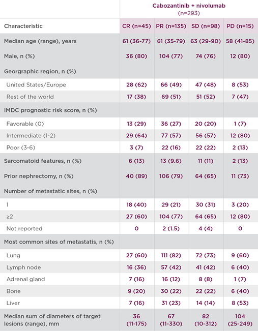
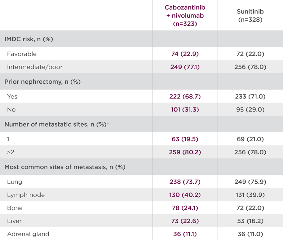
CheckMate-9ER studied a broad patient population, including those with a high burden of disease. Select baseline characteristics are shown in Table 1 for the cabozantinib + nivolumab arm.3

Table 13

Renal-Cell-Carcinoma-IMDC-Risk


Data are for tumor sites defined at baseline by the investigators according to RECIST v1.1. The number of target or nontarget lesions at baseline was not reported for 1 patient in the CABOMETYX + OPDIVO group and for 3 patients in the sunitinib group.3

Results in the CheckMate-9ER primary analysis of the intent-to-treat population (median follow-up of 18.1 months; range: 10.6-30.6 months) were as follows3:

  • Median PFS* (mPFS) was 16.6 months with cabozantinib + nivolumab (95% CI: 12.5-24.9) vs 8.3 months  with sunitinib (95% CI: 7.0-9.7) (HR=0.51; 95% CI: 0.41-0.64; P<0.0001)4,6
  • Median OS (mOS) was not reached in either arm (HR=0.60; 98.89% CI: 0.40-0.89; P=0.001)4,6
    • Pre-planned final analysis (median follow-up: 32.9 months; range: 25.4-45.4 months): mOS was 37.7 months for the combination (95% Cl: 35.5-NR) vs 34.3 months with sunitinib (95% Cl: 29.0-NR) (HR=0.70; 95% CI: 0.55-0.90)
  • ORR* was 55.7% (95% CI: 50.1-61.2) with the combination vs 27.1% with sunitinib (95% CI: 22.4-32.3; P<0.0001)4,6
    • Complete response (CR) was 8% (n=26/323) for the combination arm vs 4.6% (n=15/328) with sunitinib
    • Partial response (PR) was 48% (n=154/323) for the combination arm vs
      23% (n=74/328) with sunitinib
  • Serious adverse reactions occurred in 48% of patients receiving cabozantinib + nivolumab (n=320). The most frequent serious adverse reactions reported in ≥2% of patients were diarrhea, pneumonia, pneumonitis, pulmonary embolism, urinary tract infection, and hyponatremia. Fatal intestinal perforations occurred in 3 (0.9%) patients4
  • The most common adverse reactions (≥20%) were diarrhea (64%), fatigue (51%), hepatotoxicity (44%), palmar‐plantar erythrodysesthesia (PPE) (40%), stomatitis (37%), rash (36%), hypertension (36%), hypothyroidism (34%), musculoskeletal pain (33%), decreased appetite (28%), nausea (27%), dysgeusia (24%), abdominal pain (22%), upper respiratory tract infection (20%), and cough (20%)4

The results of the CheckMate-9ER 5-year follow-up analysis (median follow-up of 67.6 months; range 60.2-80.2 months) were as follows. No formal statistical testing was conducted at the time of the updated analysis5:

  • mPFS* was 16.4 months with the combination (95% CI: 12.5-19.3) vs 8.3 months with sunitinib (95% CI: 7.0-9.7) (HR=0.58; 95% CI: 0.49-0.70)
  • mOS was 46.5 months with the combination (95% CI: 40.6-53.8) vs 35.5 months with sunitinib (95% CI: 29.2-42.8) (HR=0.79; 95% CI: 0.65-0.96)
  • ORR* was 55.7% (95% CI: 50.1-61.2) for the combination arm vs 27.4% with sunitinib (95% CI: 22.7-32.6)
    • CR was 13.9% (n=45/323) for the combination arm vs 4.6% (n=15/328)
      with sunitinib
    • PR was 41.8% (n=135/323) for the combination arm vs 22.9% (n=75/328)
      with sunitinib
  • Safety and tolerability were similar to previous follow-ups

*Assessed by BICR.4

To further explore long-term data from the CheckMate-9ER trial, post hoc analyses were conducted to evaluate the baseline characteristics of patients who achieved CR, assess depth of response (DepOR) among those patients with PR, and examine the duration of response in patients with CR or PR.1

Depth and durability of response in patients treated with cabozantinib + nivolumab
The exploratory, post hoc analyses of the 5-year CheckMate-9ER data evaluated depth and durability of response in patients randomized to cabozantinib + nivolumab who were alive 6 months after randomization and who had available data for best overall response (BOR) and sum of diameters of target lesions.1

  • Of the 323 patients randomized to cabozantinib + nivolumab, 293 were alive at the
    6-month landmark
  • Patients were categorized into DepOR subgroups based on BOR per BICR using the maximum tumor reduction observed at any time during the study period

Baseline demographic and disease characteristics of patients receiving cabozantinib + nivolumab by DepOR subgroup are shown in Table 2, which includes patients with characteristics associated with poor prognosis.1

Table 21,7

Characteristics-Demographics


  • Among patients who achieved CR (n=45), the median duration of response was not reached at the time of analysis (95% CI: 30.5-NE)1*
  • Among patients who achieved PR (n=135), the median duration of response was 17.3 months (95% CI: 13.8-20.7)7*
  • Median duration of treatment with cabozantinib + nivolumab was 30.4 months for patients achieving CR, over 2 years for those with PR, and 15.3 months for those with stable disease (SD)1*
  • Safety was similar across all subgroups achieving an objective response, and the most frequently reported Grade 3/4 treatment-related adverse events included hypertension (13%), diarrhea (9%), hyponatremia (8%), and PPE (8%)1

These post hoc exploratory analyses are descriptive in nature. No statistical procedure was employed. Results should be considered hypothesis generating.

*Patients who were alive at the 6-month timepoint.1,7

Discussion
In the primary analysis cabozantinib + nivolumab doubled mPFS and demonstrated superior efficacy vs sunitinib across OS and ORR.4 Building on these results, the primary results and exploratory analyses from CheckMate-9ER provide additional context to the established efficacy of cabozantinib + nivolumab in the first-line treatment of aRCC.1,4 Notably, meaningful responses were observed among patients with baseline features traditionally associated with poorer prognosis, underscoring the broad applicability of this combination.1

The median duration of response was not reached among patients achieving complete response. The durability of responses and extended treatment exposure did not appear to be associated with a disproportionate increase in toxicity.1

These findings are derived from post hoc, descriptive exploratory analyses and should be interpreted with caution. Collectively, these data help refine our understanding of which patients may derive the greatest response and further support the role of cabozantinib + nivolumab combination as a first-line therapy option in aRCC.1

Dr Manojkumar Bupathi received a fee for participating in the development of this article, and his comments reflect his opinions and are not intended to constitute medical advice for individual patients.

AXL=AXL receptor tyrosine kinase; BICR=blinded independent central review; CI=confidence interval; HR=hazard ratio; IMDC=International Metastatic RCC Database Consortium; MET=hepatocyte growth factor receptor; NE=not estimable; NR=not reported; ORR=objective response rate; OS=overall survival; PD=progressive disease; PFS=progression-free survival; RECIST=Response Evaluation Criteria in Solid Tumors; VEGFR=vascular endothelial growth factor receptor.

INDICATION
CABOMETYX® (cabozantinib), in combination with nivolumab, is indicated for the first-line treatment of patients with advanced renal cell carcinoma (RCC).

IMPORTANT SAFETY INFORMATION

WARNINGS AND PRECAUTIONS

Hemorrhage: CABOMETYX can cause severe and fatal hemorrhages. The incidence of Grade 3-5 hemorrhagic events was 5% in CABOMETYX patients in RCC, HCC, and DTC studies. Discontinue CABOMETYX for Grade 3-4 hemorrhage and before surgery. Do not administer to patients who have a recent history of hemorrhage, including hemoptysis, hematemesis, or melena.

Perforations and Fistulas: Fistulas, including fatal cases, and gastrointestinal (GI) perforations, including fatal cases, each occurred in 1% of CABOMETYX patients. Monitor for signs and symptoms, and discontinue CABOMETYX in patients with Grade 4 fistulas or GI perforation.

Thromboembolic Events: CABOMETYX can cause arterial or venous thromboembolic events. Venous thromboembolism occurred in 7% (including 4% pulmonary embolism) and arterial thromboembolism in 2% of CABOMETYX patients. Fatal thrombotic events have occurred. Discontinue CABOMETYX in patients who develop an acute myocardial infarction or serious arterial or venous thromboembolic events.

Hypertension and Hypertensive Crisis: CABOMETYX can cause hypertension, including hypertensive crisis. Hypertension was reported in 37% (16% Grade 3 and <1% Grade 4) of CABOMETYX patients. In CABINET (n=195), hypertension occurred in 65% (26% Grade 3) of CABOMETYX patients. Do not initiate CABOMETYX in patients with uncontrolled hypertension. Monitor blood pressure regularly during CABOMETYX treatment. Withhold CABOMETYX for hypertension that is not adequately controlled; when controlled, resume at a reduced dose. Permanently discontinue CABOMETYX for severe hypertension that cannot be controlled with antihypertensive therapy or for hypertensive crisis.

Cardiac Failure: CABOMETYX can cause severe and fatal cardiac failure. Cardiac failure occurred in 0.5% of patients treated with CABOMETYX as a single agent, including fatal cardiac failure in 0.1% of patients. Consider baseline and periodic evaluations of left ventricular ejection fraction. Monitor for signs and symptoms of cardiovascular events. Withhold and resume at a reduced dose upon recovery or permanently discontinue depending on the severity.

Diarrhea: CABOMETYX can cause diarrhea and it occurred in 62% (10% Grade 3) of treated patients. Monitor and manage patients using antidiarrheals as indicated. Withhold CABOMETYX until improvement to ≤ Grade 1; resume at a reduced dose.

Palmar-Plantar Erythrodysesthesia (PPE): CABOMETYX can cause PPE and it occurred in 45% of treated patients (13% Grade 3). Withhold CABOMETYX until PPE resolves or decreases to
Grade 1 and resume at a reduced dose for intolerable Grade 2 PPE or Grade 3 PPE.

Hepatotoxicity: CABOMETYX in combination with nivolumab in RCC can cause hepatic toxicity with higher frequencies of Grades 3 and 4 ALT and AST elevations compared to CABOMETYX alone. With the combination of CABOMETYX and nivolumab, Grades 3 and 4 increased ALT or AST were seen in 11% of patients. Monitor liver enzymes before initiation of treatment and periodically. Consider more frequent monitoring as compared to when the drugs are administered as single agents. Consider withholding CABOMETYX and/or nivolumab, initiating corticosteroid therapy, and/or permanently discontinuing the combination for severe or
life-threatening hepatotoxicity.

Adrenal Insufficiency: CABOMETYX in combination with nivolumab can cause primary or secondary adrenal insufficiency. Adrenal insufficiency occurred in 4.7% (15/320) of patients with RCC who received CABOMETYX with nivolumab, including Grade 3 (2.2%), and Grade 2 (1.9%) adverse reactions. Withhold CABOMETYX and/or nivolumab and resume CABOMETYX at a reduced dose depending on severity.

Proteinuria: Proteinuria was observed in 8% of CABOMETYX patients. Monitor urine protein regularly during CABOMETYX treatment. For Grade 2 or 3 proteinuria, withhold CABOMETYX until improvement to ≤ Grade 1 proteinuria; resume CABOMETYX at a reduced dose. Discontinue CABOMETYX in patients who develop nephrotic syndrome.

Osteonecrosis of the Jaw (ONJ): CABOMETYX can cause ONJ and it occurred in <1% of treated patients. Perform an oral examination prior to CABOMETYX initiation and periodically during treatment. Advise patients regarding good oral hygiene practices. Withhold CABOMETYX for at least 3 weeks prior to scheduled dental surgery or invasive dental procedures. Withhold CABOMETYX for development of ONJ until complete resolution; resume at a reduced dose.

Impaired Wound Healing: CABOMETYX can cause impaired wound healing. Withhold CABOMETYX for at least 3 weeks prior to elective surgery. Do not administer for at least 2 weeks after major surgery and until adequate wound healing. The safety of resumption of CABOMETYX after resolution of wound healing complications has not been established.

Reversible Posterior Leukoencephalopathy Syndrome (RPLS): CABOMETYX can cause RPLS. Perform evaluation for RPLS and diagnose by characteristic finding on MRI any patient presenting with seizures, headache, visual disturbances, confusion, or altered mental function. Discontinue CABOMETYX in patients who develop RPLS.

Thyroid Dysfunction: CABOMETYX can cause thyroid dysfunction, primarily hypothyroidism, and it occurred in 19% of treated patients (0.4% Grade 3). Assess for signs of thyroid dysfunction prior to the initiation of CABOMETYX and monitor for signs and symptoms during treatment.

Hypocalcemia: CABOMETYX can cause hypocalcemia, with the highest incidence in DTC patients. Based on the safety population, hypocalcemia occurred in 13% of CABOMETYX patients (2% Grade 3 and 1% Grade 4).
Monitor blood calcium levels and replace calcium as necessary during treatment. Withhold and resume CABOMETYX at a reduced dose upon recovery or permanently discontinue CABOMETYX depending on severity.

Embryo-Fetal Toxicity: CABOMETYX can cause fetal harm. Advise pregnant women of the potential risk to a fetus and advise females of reproductive potential to use effective contraception during treatment with CABOMETYX and for 4 months after the last dose.

ADVERSE REACTIONS

The most common (≥20%) adverse reactions are:
CABOMETYX in combination with nivolumab: diarrhea, fatigue, hepatotoxicity, PPE, stomatitis, rash, hypertension, hypothyroidism, musculoskeletal pain, decreased appetite, nausea, dysgeusia, abdominal pain, cough, and upper respiratory tract infection.

DRUG INTERACTIONS

Strong CYP3A4 Inhibitors: If coadministration with strong CYP3A4 inhibitors cannot be avoided, reduce the CABOMETYX dosage. Avoid grapefruit or grapefruit juice.

Strong or Moderate CYP3A4 Inducers: If coadministration with strong or moderate CYP3A4 inducers cannot be avoided, increase the CABOMETYX dosage. Avoid St. John’s wort.

USE IN SPECIFIC POPULATIONS

Lactation: Advise women not to breastfeed during CABOMETYX treatment and for 4 months after the final dose.

Hepatic Impairment: In patients with moderate hepatic impairment, reduce the CABOMETYX dosage. Avoid CABOMETYX in patients with severe hepatic impairment.

Please see accompanying full Prescribing Information by clicking here.

You are encouraged to report negative side effects of prescription drugs to the FDA. Visit www.FDA.gov/medwatch or call 1-800-FDA-1088.

 References

  1. Suárez C, Motzer RJ, Powles T, et al. Characterization of depth and durability of response in patients with previously untreated advanced renal cell carcinoma who received cabozantinib plus nivolumab: long-term follow-up and exploratory analysis of CheckMate 9ER. Poster Presented at: American Society of Clinical Oncology Genitourinary Cancers Symposium. February 26-28, 2026.
  2. Zarrabi KK, Lanade O, Geynisman DM. Determining front-line therapeutic strategy for metastatic clear cell renal cell carcinoma. Cancers. 2022;14;4607. doi.org/10.3390/cancers14194607.
  3. Choueiri TK, Powles T, Burotto M, et al; CheckMate 9ER Investigators. Nivolumab plus cabozantinib versus sunitinib for advanced renal‐cell carcinoma. N Engl J Med. 2021;384(9):829‐841.
  4. CABOMETYX® (cabozantinib) Prescribing Information. Exelixis, Inc.
  5. Motzer RJ, Escudier B, Burotto M, et al. Final analysis of nivolumab plus cabozantinib for advanced renal cell carcinoma from the randomized phase III CheckMate 9ER trial. Ann Oncol. 2025; Published online September 23, 2025. doi:10.1016/j.annonc.2025.09.006.
  6. Powles T, Choueiri TK, Burotto M, et al. Final overall survival analysis and organ-specific target lesion assessments with 2-year follow-up in CheckMate 9ER: nivolumab plus cabozantinib versus sunitinib for patients with advanced renal cell carcinoma. Poster presented at: American Society of Clinical Oncology Genitourinary Cancers Symposium; February 17-19, 2022.
  7. Data on file. Exelixis, Inc.

©2026 Exelixis, Inc.    CA-3913    04/26
OPDIVO® and the related logo are registered trademarks of Bristol‐Myers Squibb Company.

Expanding systemic treatment options in neuroendocrine tumors

Insights from the Phase 3 CABINET trial of CABOMETYX® (cabozantinib) including lung and thymic NET

Written by Munveer Bhangoo, MD
Sponsored by Exelixis, Inc.

Neuroendocrine tumors (NETs) are often characterized as a diverse category of diseases that often have widely differing clinical behavior and outcomes.  This behavior is at least in part a reflection of the fact that neuroendocrine cells are scattered through the body. Tumors evolving from neuroendocrine cells vary considerably in terms of location, biological aggressiveness, hormone status, and somatostatin receptor status.1 Taken together, patients with this disease exhibit highly variable clinical outcomes.2  Furthermore, the varied clinical and pathologic features of NETs impact the landscape of clinical trials in this space, with a longstanding need for trials to include a population representative of the disease.3,4

Metastatic neuroendocrine tumors have historically represented an unmet need, particularly for patients experiencing disease progression. In March 2025, the FDA approval of cabozantinib (CABOMETYX®) for the treatment of adult and pediatric patients 12 years of age and older with previously treated, unresectable, locally advanced or metastatic, well-differentiated pancreatic neuroendocrine tumors (pNET) or extrapancreatic neuroendocrine tumors (epNET) marked a meaningful advancement in addressing this gap.3,5  As the only FDA-approved option for patients with previously treated NET, regardless of primary site, tumor grade, SSTR expression, or functional disease, CABOMETYX offers an approved treatment option for patients whose disease progresses on an initial therapy.5-11

Let’s review the pivotal trial data from CABINET, a Phase 3 trial enrolling a heterogeneous population with both pancreatic and extrapancreatic NET, which spanned gastrointestinal, lung, thymus, and unknown and other sites of origin.3

CABINET trial design

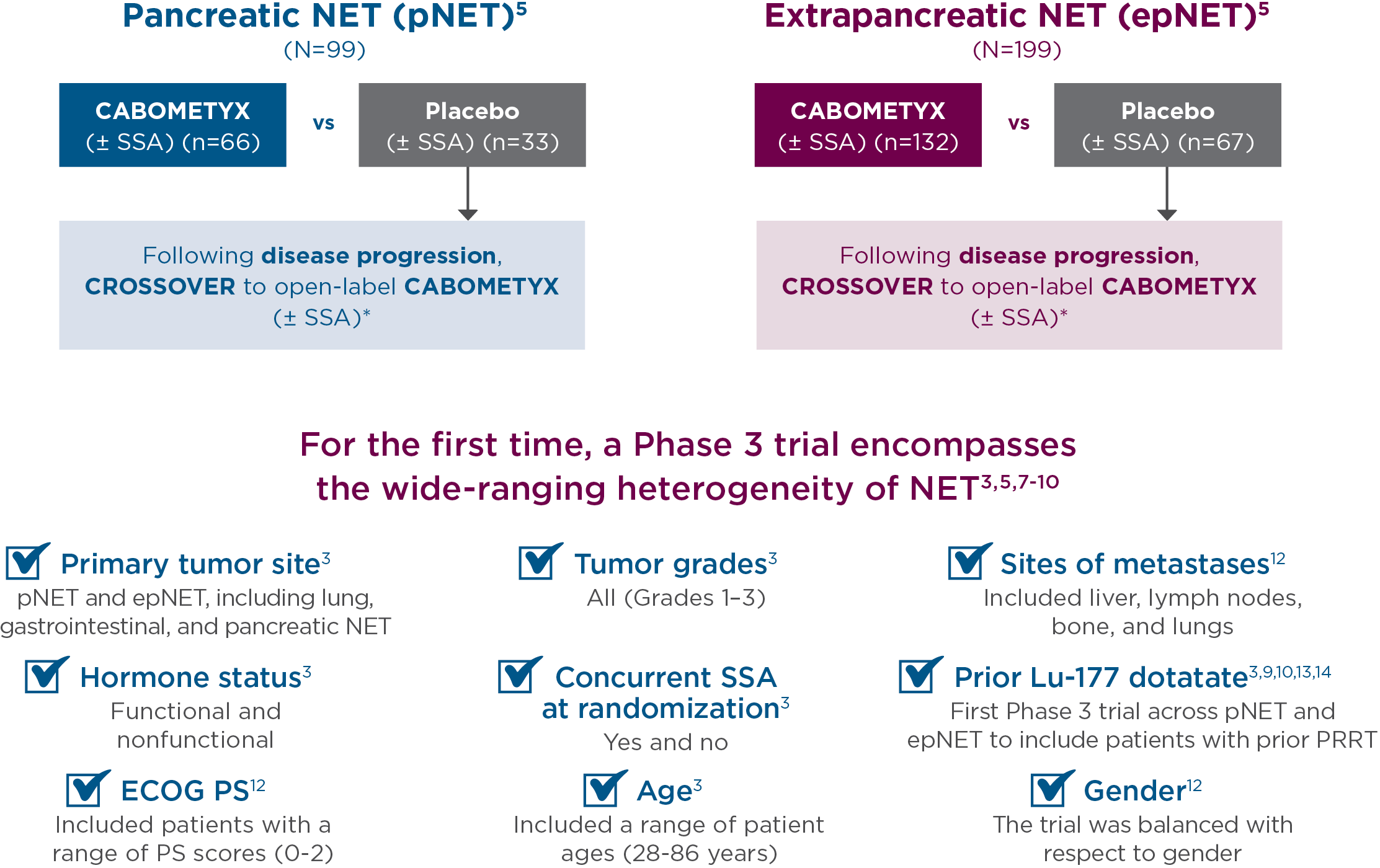
CABINET was a randomized (2:1), double-blind, placebo-controlled, Phase 3, National Cancer Institute-sponsored trial of CABOMETYX vs placebo in advanced NET patients previously treated with ≥1 FDA-approved systemic therapy, not including an SSA. CABINET enrolled 2 independent cohorts that evaluated patients with pNET (N=99) or epNET (N=199). The starting dose for CABOMETYX was 60 mg, administered orally once daily. The primary endpoint was PFS; ORR and OS were secondary endpoints.3,5


Study-Design-Exelixis-NET

*Unblinding and crossover to open-label CABOMETYX allowed after confirmation of progressive disease by real-time central radiology review.5


Primary efficacy results in CABINET

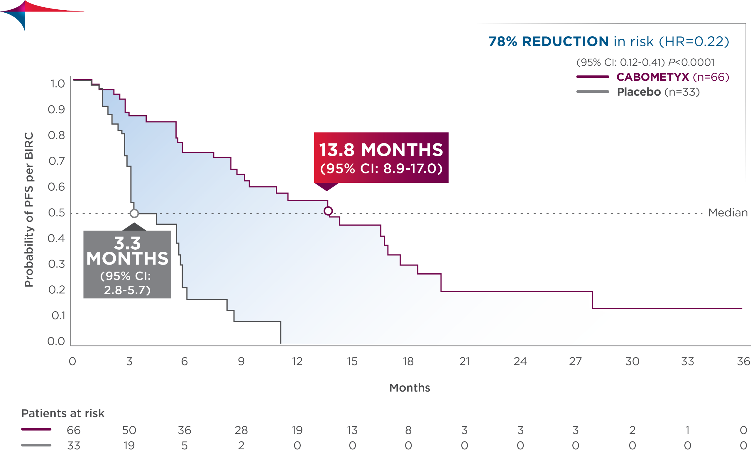
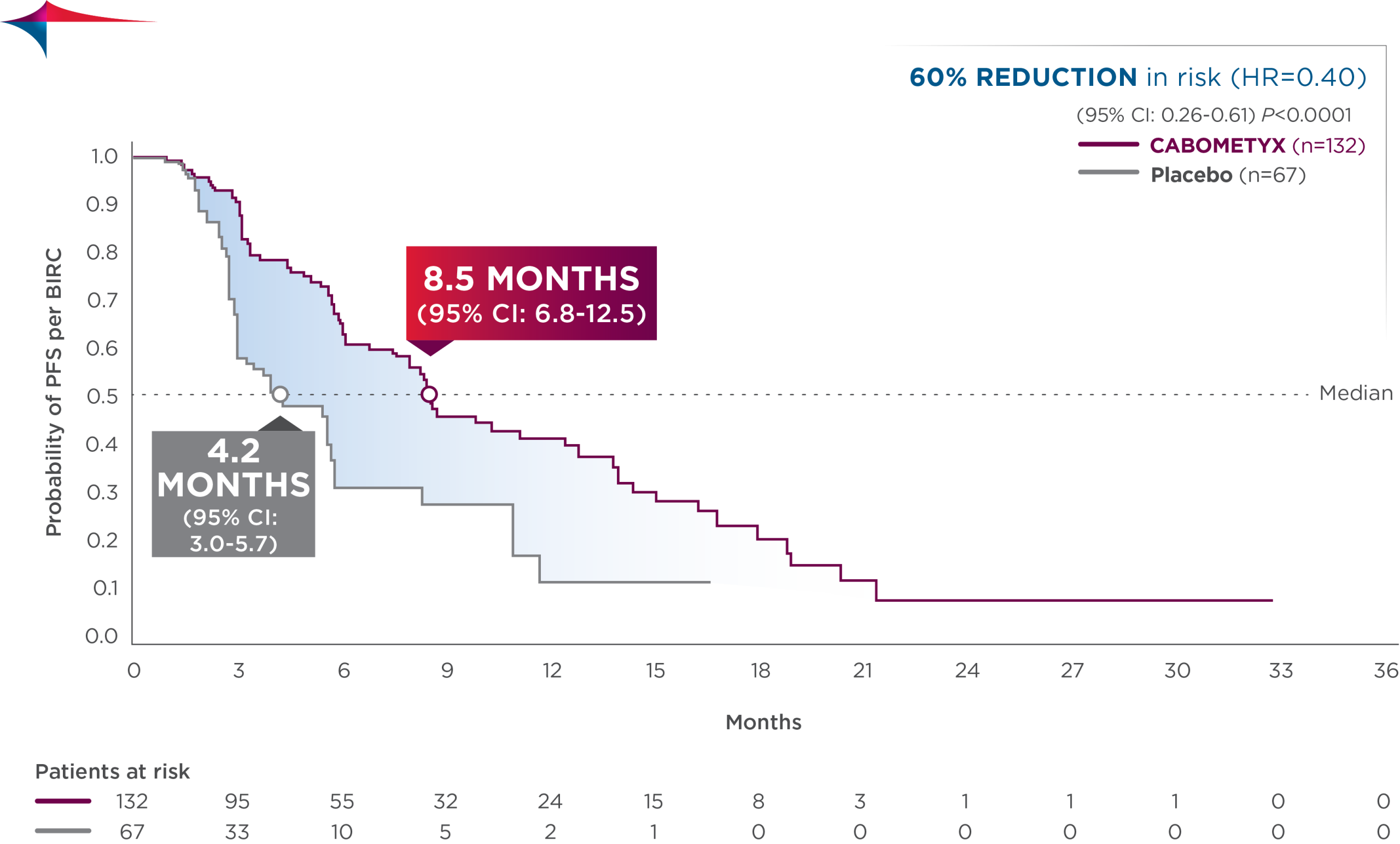
CABOMETYX quadrupled median PFS in pNET and doubled median PFS in epNET5

  • pNET: median PFS was 13.8 months (95% CI: 8.9-17.0; n=66) vs 3.3 months with placebo (95% CI: 2.8-5.7; n=33); HR=0.22 (95% CI: 0.12-0.41); P<0.0001
  • epNET: median PFS was 8.5 months (95% CI: 6.8-12.5; n=132) vs 4.2 months with placebo (95% CI: 3.0-5.7; n=67); HR=0.40 (95% CI: 0.26-0.61); P<0.0001

pNET-PFS-CABOMETYX


epNET-PFS-CABOMETYX


Exploratory subgroup analysis: PFS results in patients with lung/thymus site origin15

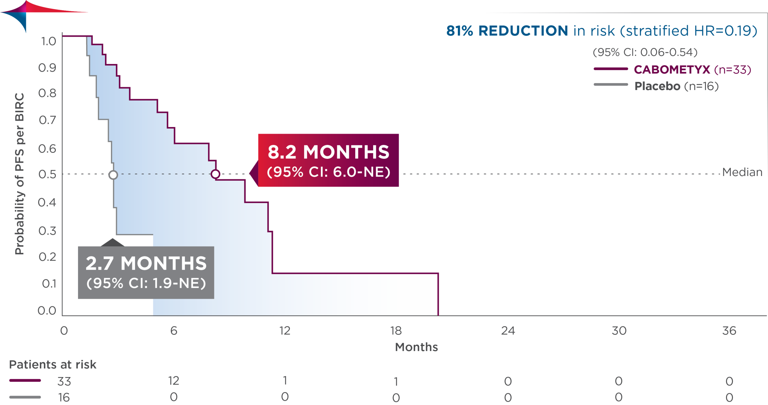
  • In the lung /thymus subgroup, median PFS was 8.2 months (95% CI: 6.0-NE; n=33) vs 2.7 months (95% CI: 1.9-NE; n=16) with placebo; HR=0.19 (95% CI: 0.06-0.54)

Lung-Thymus-Subgroup-PFS-CABOMETYX


Observed outcomes should be interpreted with caution because of the relatively small subgroup size. Subgroups were not powered to show differences between treatment arms, and results should be considered hypothesis generating.

CABINET safety results

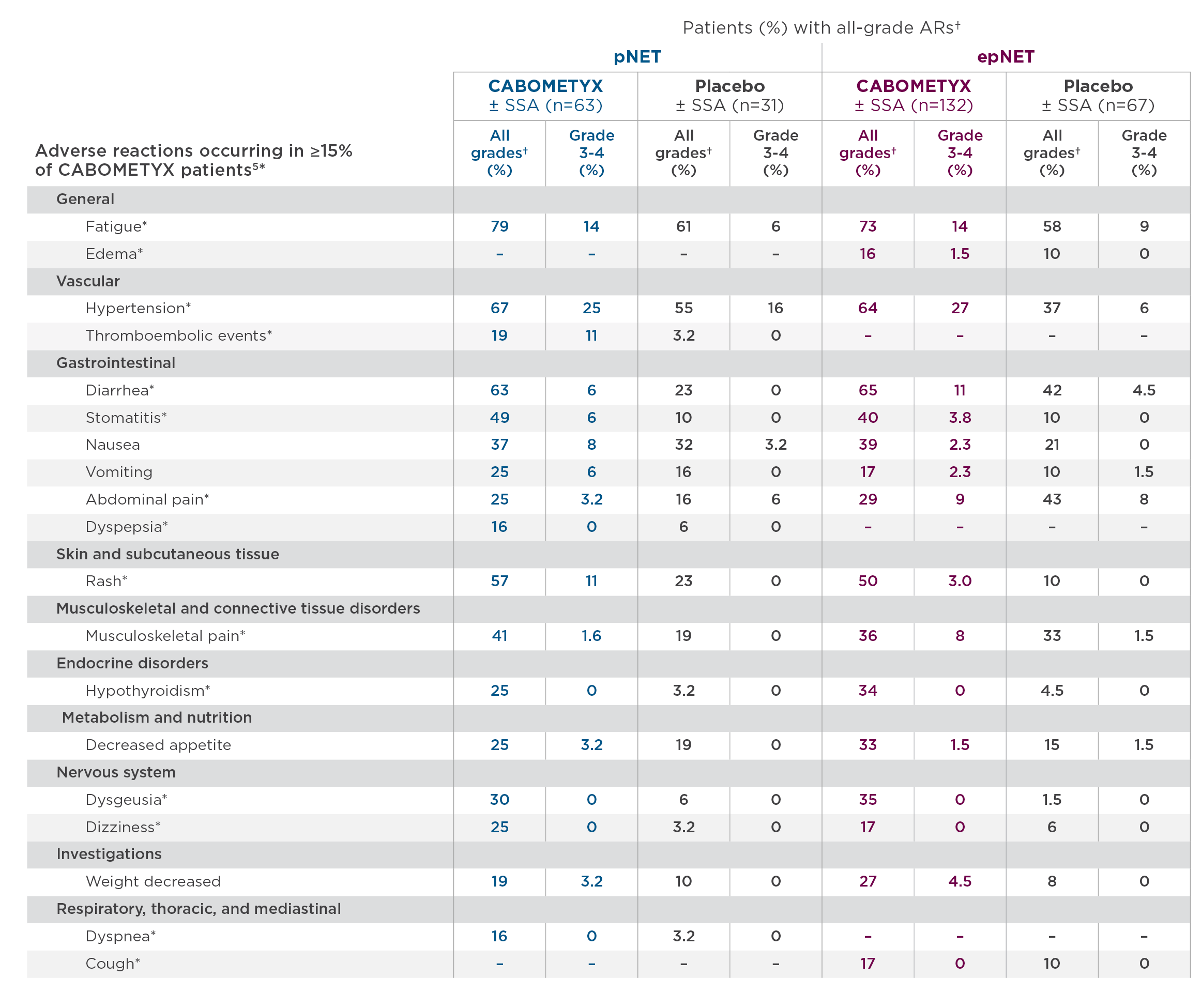
The safety profile observed in CABINET was consistent with the known CABOMETYX safety profile, and no new safety signals were identified in the trial.3

AE-CABOMETYX

  • Commonly occurring treatment-related Grade 3 and 4 adverse events in the lung/thymus subgroup included fatigue, hypertension, diarrhea, and PPE15

*These ARs are grouped terms.5 For details, please see full Prescribing Information.
NCI CTCAE Version 5.0.

CABOMETYX dosing for NET

The recommended starting dose of CABOMETYX for adult and pediatric patients 12 years of age and older and ≥40 kg is 60 mg once daily until disease progression or unacceptable toxicity (with dose reductions to 40 mg and 20 mg once daily). The recommended starting dose for pediatric patients 12 years of age and older and <40 kg is 40 mg (with dose reductions to 20 mg daily and 20 mg every other day). If previously receiving lowest dose, resume at same dose. If lowest dose not tolerated, discontinue CABOMETYX. The median average daily dose of CABOMETYX treatment was 41 mg in the pNET cohort and 43 mg in the epNET cohort.5

It is important to note that the overall efficacy results of the CABINET trial were achieved in the context of dose modifications.16

  • pNET: AR-related dose reductions occurred in 49% of patients receiving CABOMETYX vs 16% with placebo. Discontinuation due to ARs occurred in 19% of patients receiving CABOMETYX vs 10% with placebo
  • epNET: AR-related dose reductions occurred in 38% of patients receiving CABOMETYX vs 6% with placebo. Discontinuation due to ARs occurred in 28% of patients receiving CABOMETYX vs 19% receiving placebo

In summary, based on the CABINET data:

Cabozantinib became the first systemic therapy indicated for previously treated NETs regardless of origin based on the landmark Phase 3 CABINET trial.3,5,7-11 Furthermore, the broad eligibility criteria for CABINET allowed for the inclusion of patients irrespective of functional status or expression of somatostatin receptors.3

Taken together, the approval of cabozantinib represents an important advancement in the management of previously treated patients with advanced NETs across diverse sites of origin, including those with lung and thymic origin.3,5,17


Dr Bhangoo received a fee for participating in the development of this article, and his comments reflect his opinions and are not intended to constitute medical advice for individual patients

AR=adverse reaction; CI=confidence interval; DCR=disease control rate; ECOG PS=Eastern Cooperative Oncology Group performance status; epNET=extrapancreatic neuroendocrine tumor; HR=hazard ratio; NET=neuroendocrine tumor; ORR=overall response rate; OS=overall survival; NE=not estimable; PET=positron emission tomography; PFS=progression-free survival; pNET=pancreatic neuroendocrine tumor; PPE=palmar-plantar erythrodysesthesia; PRRT=peptide receptor radionuclide therapy.

INDICATIONS

CABOMETYX® (cabozantinib) is indicated for the treatment of adult and pediatric patients 12 years of age and older with previously treated, unresectable, locally advanced or metastatic, well-differentiated pancreatic neuroendocrine tumors (pNET).

CABOMETYX is indicated for the treatment of adult and pediatric patients 12 years of age and older with previously treated, unresectable, locally advanced or metastatic, well-differentiated extrapancreatic neuroendocrine tumors (epNET).

IMPORTANT SAFETY INFORMATION

WARNINGS AND PRECAUTIONS

Hemorrhage: CABOMETYX can cause severe and fatal hemorrhages. The incidence of Grade 3-5 hemorrhagic events was 5% in CABOMETYX patients in RCC, HCC, and DTC studies. Discontinue CABOMETYX for Grade 3-4 hemorrhage and before surgery. Do not administer to patients who have a recent history of hemorrhage, including hemoptysis, hematemesis, or melena.

Perforations and Fistulas: Fistulas, including fatal cases, and gastrointestinal (GI) perforations, including fatal cases, each occurred in 1% of CABOMETYX patients. Monitor for signs and symptoms, and discontinue CABOMETYX in patients with Grade 4 fistulas or GI perforation.

Thromboembolic Events: CABOMETYX can cause arterial or venous thromboembolic events. Venous thromboembolism occurred in 7% (including 4% pulmonary embolism) and arterial thromboembolism in 2% of CABOMETYX patients. Fatal thrombotic events have occurred. Discontinue CABOMETYX in patients who develop an acute myocardial infarction or serious arterial or venous thromboembolic events.

Hypertension and Hypertensive Crisis: CABOMETYX can cause hypertension, including hypertensive crisis. Hypertension was reported in 37% (16% Grade 3 and <1% Grade 4) of CABOMETYX patients. In CABINET (n=195), hypertension occurred in 65% (26% Grade 3) of CABOMETYX patients. Do not initiate CABOMETYX in patients with uncontrolled hypertension. Monitor blood pressure regularly during CABOMETYX treatment. Withhold CABOMETYX for hypertension that is not adequately controlled; when controlled, resume at a reduced dose. Permanently discontinue CABOMETYX for severe hypertension that cannot be controlled with antihypertensive therapy or for hypertensive crisis.

Cardiac Failure: CABOMETYX can cause severe and fatal cardiac failure. Cardiac failure occurred in 0.5% of patients treated with CABOMETYX as a single agent, including fatal cardiac failure in 0.1% of patients. Consider baseline and periodic evaluations of left ventricular ejection fraction. Monitor for signs and symptoms of cardiovascular events. Withhold and resume at a reduced dose upon recovery or permanently discontinue depending on the severity.

Diarrhea: CABOMETYX can cause diarrhea and it occurred in 62% (10% Grade 3) of treated patients. Monitor and manage patients using antidiarrheals as indicated. Withhold CABOMETYX until improvement to ≤ Grade 1; resume at a reduced dose.

Palmar-Plantar Erythrodysesthesia (PPE): CABOMETYX can cause PPE and it occurred in 45% of treated patients (13% Grade 3). Withhold CABOMETYX until PPE resolves or decreases to Grade 1 and resume at a reduced dose for intolerable Grade 2 PPE or Grade 3 PPE.

Proteinuria: Proteinuria was observed in 8% of CABOMETYX patients. Monitor urine protein regularly during CABOMETYX treatment. For Grade 2 or 3 proteinuria, withhold CABOMETYX until improvement to ≤ Grade 1 proteinuria; resume CABOMETYX at a reduced dose. Discontinue CABOMETYX in patients who develop nephrotic syndrome.

Osteonecrosis of the Jaw (ONJ): CABOMETYX can cause ONJ and it occurred in <1% of treated patients. Perform an oral examination prior to CABOMETYX initiation and periodically during treatment. Advise patients regarding good oral hygiene practices. Withhold CABOMETYX for at least 3 weeks prior to scheduled dental surgery or invasive dental procedures. Withhold CABOMETYX for development of ONJ until complete resolution; resume at a reduced dose.

Impaired Wound Healing: CABOMETYX can cause impaired wound healing. Withhold CABOMETYX for at least 3 weeks prior to elective surgery. Do not administer for at least 2 weeks after major surgery and until adequate wound healing. The safety of resumption of CABOMETYX after resolution of wound healing complications has not been established.

Reversible Posterior Leukoencephalopathy Syndrome (RPLS): CABOMETYX can cause RPLS. Perform evaluation for RPLS and diagnose by characteristic finding on MRI any patient presenting with seizures, headache, visual disturbances, confusion, or altered mental function. Discontinue CABOMETYX in patients who develop RPLS.

Thyroid Dysfunction: CABOMETYX can cause thyroid dysfunction, primarily hypothyroidism, and it occurred in 19% of treated patients (0.4% Grade 3). Assess for signs of thyroid dysfunction prior to the initiation of CABOMETYX and monitor for signs and symptoms during treatment.

Hypocalcemia: CABOMETYX can cause hypocalcemia, with the highest incidence in DTC patients. Based on the safety population, hypocalcemia occurred in 13% of CABOMETYX patients (2% Grade 3 and 1% Grade 4). Monitor blood calcium levels and replace calcium as necessary during treatment. Withhold and resume CABOMETYX at a reduced dose upon recovery or permanently discontinue CABOMETYX depending on severity.

Embryo-Fetal Toxicity: CABOMETYX can cause fetal harm. Advise pregnant women of the potential risk to a fetus and advise females of reproductive potential to use effective contraception during treatment with CABOMETYX and for 4 months after the last dose.

ADVERSE REACTIONS The most common (≥20%) adverse reactions are: CABOMETYX as a single agent: diarrhea, fatigue, PPE, decreased appetite, hypertension, nausea, vomiting, weight decreased, and constipation.

DRUG INTERACTIONS

Strong CYP3A4 Inhibitors: If coadministration with strong CYP3A4 inhibitors cannot be avoided, reduce the CABOMETYX dosage. Avoid grapefruit or grapefruit juice.

Strong or Moderate CYP3A4 Inducers: If coadministration with strong or moderate CYP3A4 inducers cannot be avoided, increase the CABOMETYX dosage. Avoid St. John’s wort.

USE IN SPECIFIC POPULATIONS

Lactation: Advise women not to breastfeed during CABOMETYX treatment and for 4 months after the final dose.

Hepatic Impairment: In patients with moderate hepatic impairment, reduce the CABOMETYX dosage. Avoid CABOMETYX in patients with severe hepatic impairment.

Pediatric Use: Physeal widening has been observed in children with open growth plates when treated with CABOMETYX. Physeal and longitudinal growth monitoring is recommended in children (12 years and older) with open growth plates. Consider interrupting or discontinuing CABOMETYX if abnormalities occur. The safety and effectiveness of CABOMETYX in pediatric patients less than 12 years of age have not been established.

Please see accompanying full Prescribing Information by clicking here.

You are encouraged to report negative side effects of prescription drugs to the FDA. Visit www.FDA.gov/medwatch or call 1-800-FDA-1088.

References:
1.
Sultana Q, Kar J, Verma A, et al. A comprehensive review on neuroendocrine neoplasms: presentation, pathophysiology and management. J Clin Med. 2023;12(15):5138
2. Dasari A, Wallace K, Halperin DM, et al. Epidemiology of neuroendocrine neoplasms in the US. JAMA Netw Open. 2025;8(6):e2515798. doi:10.1001/jamanetworkopen.2025.15798.
3. Chan JA, Geyer S, Zemla T, et al. Phase 3 trial of cabozantinib to treat advanced neuroendocrine tumors. N Engl J Med. 2024; Published online September 16, 2024. doi:10.1056/NEJMoa2403991.
4. Yang K, Li J, Cheng Y, Bai C. Evolving landscape of clinical trials in gastroenteropancreatic neuroendocrine neoplasms in the past two decades. Endocr Connect. 2023;12(4): e220441. doi:10.1530/EC-22-0441.
5. CABOMETYX® (cabozantinib) Prescribing Information. Exelixis, Inc.
6. Bidani K, Marinovic AG, Moond V, Harne P, Broder A, Thosani N. Treatment of pancreatic neuroendocrine tumors: beyond traditional surgery and targeted therapy. J Clin Med. 2025;14(10):3389. doi:10.3390/jcm14103389.
7. LUTATHERA® (lutetium Lu-177 dotatate) Prescribing Information. Novartis Pharmaceuticals Corporation.
8. AFINITOR® (everolimus) Prescribing Information. Novartis Pharmaceuticals Corporation.
9. SUTENT® (sunitinib malate) Prescribing Information. Pfizer, Inc.
10. SOMATULINE® DEPOT (lanreotide) Prescribing Information. Ipsen Pharma Biotech.
11. SANDOSTATIN® LAR DEPOT (octreotide acetate) Prescribing Information. Novartis Pharmaceuticals Corporation.
12. Chan JA, Geyer S, Zemla T, et al. Phase 3 trial of cabozantinib in previously treated advanced neuroendocrine tumors [supplementary appendix]. N Engl J Med. 2024; Published online September 16, 2024. doi:10.1056/ NEJMoa2403991.
13. US Food and Drug Administration. FDA approves new treatment for certain digestive tract cancers. January 26, 2018. Accessed September 5, 2024. https://www.fda.gov/news-events/press-announcements/fda-approves-new-treatment-certain-digestive-tract-cancers. 14. Yao JC, Shah MH, Ito T, et al. Everolimus for advanced pancreatic neuroendocrine tumors. N Engl J Med. 2011;364(6):514-523.
15. Wolin EM, Zemla T, Strosberg JR, et al. Efficacy and safety of cabozantinib for advanced lung and thymus neuroendocrine tumors after progression on prior therapy: subgroup analysis of phase 3 CABINET trial (Alliance A021602). Poster presented at European Society for Medical Oncology Congress; October 17-21, 2025.
16. Data on file. Exelixis, Inc.
17. Oronsky N, Ma PC, Morgensztern D, Carter CA. Nothing but NET: a review of neuroendocrine tumors and carcinomas. Neoplasia. 2017;19(12):991-1002.

©2026 Exelixis, Inc.      CA‐3900     04/26

Extrapulmonary Neuroendocrine Carcinoma: Clinical Overview and Advances in DLL3 Targeted Therapy

Written by: Dr. Eric Lander
Sponsored by Boehringer Ingelheim

Extrapulmonary neuroendocrine carcinomas (EP-NECs) are rare and phenotypically aggressive malignancies arising from neuroendocrine cells. While EP-NECs are currently managed with conventional chemotherapy in most cases, numerous therapies are in development which may show promise to improve disease management and prognosis for patients.

EP-NECs originate from neuroendocrine cells located in many different organs, most commonly arising from the GI tract or pancreas, followed by genitourinary tract and gynecologic organs [1]. NECs are often confused with neuroendocrine tumors (NETs). Though both NETs and NECs arise from epithelial neuroendocrine cells expressing pan-cytokeratin, synaptophysin, and Chromogranin A, by definition NETs are well-differentiated while NECs are poorly differentiated. Though NETs can be defined as grades 1-3, they are more commonly grade 1-2 (Ki-67 <20%); NECs must be grade 3 (Ki-67 ≥20% and/or mitotic count >20 per mm2), and the Ki-67 usually exceeds 50%. The remainder of this article will focus on EP-NECs and will not include discussion about grade 3 NETs. Please reference the NCCN Guidelines or the Expert Consensus Practice Recommendations of the North American Neuroendocrine Tumor Society (NANETS) to learn about management strategies for G3 NETs [2].

EP-NECs most commonly result from TP53 and RB1 inactivation, similar to small cell lung neuroendocrine carcinoma (SCLC), though EP-NECs often contribute their own unique genetic mutational background (e.g. BRAF, KRAS, PIK3CA, APC, etc.) based on their site of origin, unlike most SCLC cases. If the primary site of EP-NEC origin is unknown, as occurs in up to one third of cases, encouraging pathology to perform transcription factor IHC can facilitate a site of origin assignment. Certain transcription factors (in parentheses) are unique to each organ: midgut (CDX2); pancreas (PAX6, PAX8, islet 1, or PR); rectum (SATB2); lung (OTP, TTF-1). Delineating site of origin is of particular importance as EP-NEC may be treated according to its primary site of origin at time of relapse following platinum-based chemotherapy.

Since EP-NECs are aggressive, high-grade carcinomas, patients most commonly have metastatic disease at the time of presentation. Many patients initially present for the first time to the hospital because some symptom of their disease, such as severe pain or fracture in the case of bone metastases, necessitated their presentation to the emergency room. Initial workup following tissue diagnosis should consist of imaging of the chest/abdomen/pelvis with CT or FDG-PET/CT imaging. Notably, high grade NECs have lower somatostatin receptor (SSTR) expression than NETs; therefore, FDG is preferred over SSTR-PET radiotracers [3, 4]. For EP-NECs, the incidence of brain metastases is less than 2%; thus, brain MRI should only be considered at time of diagnosis in cases of high disease burden or in symptomatic patients [5].

For molecular workup, since many EP-NECs can harbor mutations in BRAF (particularly in colorectal EP-NECs) and tumor agnostic indications for other therapies exist, NGS testing may be considered. Mismatch repair (MMR) testing or MSI testing is also recommended since 10% of NECs are deficient MMR, opening the door to immunotherapies as therapeutic options. Delta-like ligand 3 (DLL3) is an emerging target in EP-NEC; reserving tissue for DLL3 IHC is recommended in cases where patients may enroll in a clinical trial investigating a drug targeting DLL3 – which will be discussed later.

For the management of localized EP-NEC, discussion at tumor board is recommended to provide a multidisciplinary treatment approach. Data surrounding the long-term curative potential of surgery is mixed based on the tumor site of origin when surgery is often invasive, and patients remain at high risk of metastatic disease recurrence. For this reason, neoadjuvant or adjuvant platinum-based chemotherapy may be paired with surgery. Many experts will favor neoadjuvant platinum/etoposide chemotherapy to test the biology of the disease and decrease theoretical risk of micro-metastasis prior to surgery. However, many patients will present to medical oncology following tumor resection, in which case adjuvant chemotherapy may be discussed with eligible patients. Otherwise, definitive chemoradiation for organ preservation may be considered with platinum plus etoposide as the recommended radiosensitizing agents. The accruing French NEONEC trial will prospectively test neoadjuvant chemotherapy followed by surgery or chemoradiation in patients to hopefully offer clarity regarding the optimal multidisciplinary approach [6].

In the case of metastatic EP-NECs, the treatment paradigm initially parallels that of SCLC. Enrollment in clinical trial when available or platinum plus etoposide for four to six cycles remains the current first-line standard-of-care. Unlike SCLC, atezolizumab is not written into the NCCN guidelines for EP-NEC. EP-NEC patients were not included in the IMpower133 trial, and a subsequent retrospective study of a small EP-NEC patient cohort did not demonstrate a PFS or OS benefit of adding atezolizumab to platinum-based chemotherapy [7]. Larger patient numbers in a prospective trial are likely required to detect a benefit of atezolizumab—an ongoing phase II/III SWOG trial is investigating platinum/etoposide with or without atezolizumab to address this evidence gap [8].

Most patients will achieve significant initial tumor shrinkage or disease control in response to carboplatin or cisplatin plus etoposide, especially if Ki-67 ≥ 55%, but the tumor response is not durable in most cases, and tumors are less responsive to chemotherapy upon disease progression. There is currently no standard second- or third-line treatment option for EP-NEC. When assessing patients’ treatment goals and performance status, best supportive care with hospice is a very reasonable approach in light of EP-NEC’s generally poor prognosis upon time of disease relapse.

When second-line and beyond therapy lines are being considered, enrollment in clinical trial is the preferred option for eligible patients. If patients experienced a durable response lasting at least 6 months following first-line platinum/etoposide, rechallenge may be considered. Among patients with gastrointestinal and pancreatic EP-NECs, second-line treatment with FOLFIRI has the most prospective data and lends a 6-month overall survival rate of 60% [9], while gynecologic EP-NEC has data for topotecan, taxanes, single agent irinotecan, or the combination of topotecan, paclitaxel, and bevacizumab that provided an 8-month median PFS in a small retrospective cohort [10]. For patients with dMMR/MSI-H or TMB-High disease, ipilimumab/nivolumab or pembrolizumab may be considered where dual checkpoint inhibition potentially yields a higher response rate [11]. For patients with BRAF V600E mutations, a STAR trial through SCRI is available to open at most US Oncology practices employing BRAF/MEK inhibition with dabrafenib/trametinib and includes patients with EP-NEC [12].

The most promising emerging therapies for EP-NEC remain those in clinical trials targeting DLL3—this assertion is based on extrapolation of promising data from the DeLLphi trials using tarlatamab in SCLC, and initial results investigating obrixtamig in SCLC and EP-NEC. Both tarlatamab and obrixtamig are DLL3/CD3 bispecific T-cell engagers. While DLL3 is expressed in approximately 90% of SCLC, rates of DLL3 expression in EP-NEC are lower [13]. Despite this, most patients with negative DLL3 expression in the DeLLphi-301 trial employing tarlatamab in refractory SCLC still experienced disease control with tarlatamab monotherapy [14]. Emerging therapeutics targeting DLL3 are mostly either DLL3/CD3 bispecific T-cell engagers or DLL3-targeting antibody-drug conjugates.

There are several clinical trials investigating DLL3/CD3 bispecific T-cell engagers and DLL3 antibody-drug conjugates in EP-NEC patients. At the time of writing, three different phase I studies are open and actively recruiting through US Oncology Network practices that include patients with EP-NEC, all of which require tissue for DLL3 IHC testing [15, 16, 17]. Among investigational DLL3/CD3 bispecific agents, Boehringer Ingelheim’s obrixtamig has shown promising results.  Data presented in 2025 from the phase I dose-escalation trial of obrixtamig showed that heavily-pretreated EP-NEC patients with high DLL3 expression had an overall response rate of 40% and duration of response of 7.9 months [18]. While not open in the US Oncology Network, the phase II DAREON-5 trial with obrixtamig is testing two different doses and includes patients with relapsed EP-NEC [19]. The results of ongoing obrixtamig trials will be important to follow and could potentially alter our future therapeutic approach to EP-NEC.

Standard-of-care options in EP-NEC do not yield survival much past one year in most patients. However, for the first time in decades, numerous emerging therapeutic options afford hope to significantly improve the treatment tolerability and prognosis for patients with this aggressive disease.

References:

  1. Dasari A, Mehta K, Byers LA, Sorbye H, Yao JC. Comparative study of lung and extrapulmonary poorly differentiated neuroendocrine carcinomas: A SEER database analysis of 162,983 cases. Cancer. 2018;124(4):807-815. doi:10.1002/cncr.31124.
  2. Eads JR, Halfdanarson TR, Asmis T, et al. Expert Consensus Practice Recommendations of the North American Neuroendocrine Tumor Society for the management of high grade gastroenteropancreatic and gynecologic neuroendocrine neoplasms. Endocr Relat Cancer. 2023;30(8):e220206. Published 2023 Jul 11. doi:10.1530/ERC-22-0206.
  3. Tomimaru Y, Eguchi H, Tatsumi M, et al. Clinical utility of 2-[(18)F] fluoro-2-deoxy-D-glucose positron emission tomography in predicting World Health Organization grade in pancreatic neuroendocrine tumors. Surgery. 2015;157(2):269-276. doi:10.1016/j.surg.2014.09.011.
  4. Majala S, Seppänen H, Kemppainen J, et al. Prediction of the aggressiveness of non-functional pancreatic neuroendocrine tumors based on the dual-tracer PET/CT. EJNMMI Res. 2019;9(1):116. Published 2019 Dec 23. doi:10.1186/s13550-019-0585-7.
  5. Alese OB, Jiang R, Shaib W, et al. High-Grade Gastrointestinal Neuroendocrine Carcinoma Management and Outcomes: A National Cancer Database Study. Oncologist. 2019;24(7):911-920. doi:10.1634/theoncologist.2018-0382.
  6. Efficacy of neoadjuvant chemotherapy in terms of DFS in patients with locally advanced, poorly differentiated digestive neuroendocrine carcinomas (NEONEC). ClinicalTrials.gov identifier NCT04268121. Updated 2025. Accessed March 9, 2026. https://clinicaltrials.gov/study/NCT04268121
  7. Ho IW, Chiang NJ, Lai JI, et al. Efficacy of atezolizumab combined with platinum and etoposide in the treatment of extrapulmonary neuroendocrine carcinoma. Oncologist. 2025;30(3):oyae372. doi:10.1093/oncolo/oyae372.
  8. Evaluating the addition of the immunotherapy drug atezolizumab to standard chemotherapy treatment for advanced or metastatic neuroendocrine carcinomas that originate outside the lung (SWOG S2012). ClinicalTrials.gov identifier NCT05058651. Updated 2026. Accessed March 9, 2026. https://clinicaltrials.gov/study/NCT05058651
  9. Walter T, Lievre A, Coriat R, et al. Bevacizumab plus FOLFIRI after failure of platinum-etoposide first-line chemotherapy in patients with advanced neuroendocrine carcinoma (PRODIGE 41-BEVANEC): a randomised, multicentre, non-comparative, open-label, phase 2 trial. Lancet Oncol. 2023;24(3):297-306. doi:10.1016/S1470-2045(23)00001-3.
  10. Frumovitz M, Munsell MF, Burzawa JK, et al. Combination therapy with topotecan, paclitaxel, and bevacizumab improves progression-free survival in recurrent small cell neuroendocrine carcinoma of the cervix. Gynecol Oncol. 2017;144(1):46-50. doi:10.1016/j.ygyno.2016.10.040.
  11. Patel SP, Mayerson E, Chae YK, et al. A phase II basket trial of Dual Anti-CTLA-4 and Anti-PD-1 Blockade in Rare Tumors (DART) SWOG S1609: High-grade neuroendocrine neoplasm cohort. Cancer. 2021;127(17):3194-3201. doi:10.1002/cncr.33591.
  12. ClinicalTrials.gov. Clinical study to further evaluate the efficacy of dabrafenib plus trametinib in patients with rare BRAF V600E mutation-positive unresectable or metastatic solid tumors. Identifier NCT05868629. Updated 2025. Accessed March 9, 2026. https://clinicaltrials.gov/study/NCT05868629
  13. Serrano AG, Rocha P, Freitas Lima C, et al. Delta-like ligand 3 (DLL3) landscape in pulmonary and extra-pulmonary neuroendocrine neoplasms. NPJ Precis Oncol. 2024;8(1):268. Published 2024 Nov 19. doi:10.1038/s41698-024-00739-y.
  14. Ahn MJ, Cho BC, Felip E, et al. Tarlatamab for Patients with Previously Treated Small-Cell Lung Cancer. N Engl J Med. 2023;389(22):2063-2075. doi:10.1056/NEJMoa2307980.
  15. ClinicalTrials.gov. A study of Peluntamig (PT217) in patients with neuroendocrine carcinomas expressing DLL3 (the SKYBRIDGE study). Identifier NCT05652686. Updated 2025. Accessed March 9, 2026. https://clinicaltrials.gov/study/NCT05652686
  16. ClinicalTrials.gov. A study of IDE849 in patients with DLL3 expressing tumors including small cell lung cancer. Identifier NCT07174583. Updated 2026. Accessed March 9, 2026. https://clinicaltrials.gov/study/NCT07174583
  17. ClinicalTrials.gov. A Phase Ib/II, open-label, multi-center study of ZL-1310 in participants with selected solid tumors. Identifier NCT06885281. Updated 2026. Accessed March 9, 2026. https://clinicaltrials.gov/study/NCT06885281
  18. Capdevila J, Gambardella V, Kuboki Y, et al. Efficacy and safety of the DLL3/CD3 T-cell engager obrixtamig in patients with extrapulmonary neuroendocrine carcinomas with high or low DLL3 expression: Results from an ongoing phase I trial. J Clin Oncol. 2025;43(16_suppl):3004. doi: 10.1200/JCO.2025.43.16_suppl.3004.
  19. ClinicalTrials.gov. DAREON-5: A study to test whether different doses of BI 764532 help people with small cell lung cancer or other neuroendocrine cancers. Identifier NCT05882058. Updated 2026. Accessed March 9, 2026. https://clinicaltrials.gov/study/NCT05882058

Expert Perspectives on MRD Testing in Multiple Myeloma

Learn how leading oncologists use MRD to inform treatment strategy and predict relapse risk

Written by: Dr. Gary Simmons & Dr. Kashif Ali
This educational opportunity is sponsored by Adaptive Biotechnologies

Measurable residual disease (MRD) testing has become a valuable tool across the multiple myeloma disease continuum, offering unprecedented insight into disease burden, treatment response, and relapse risk.  NCCN guidelines define MRD negativity as the absence of clonal plasma cells by next generation flow cytometry or next generation sequencing (NGS), at a sensitivity of at least 1 in 10-5 cells, and recommend assessing MRD status after induction, post-transplant, post-consolidation and during maintenance therapy.1  MRD results are shaping key decisions ranging from the role and timing of autologous stem cell transplant to strategies for monitoring and treatment adjustment.  Notably, MRD may be measured from bone marrow or peripheral blood, with data indicating that blood-based testing complements – but does not replace – bone marrow-based testing.2  In this dual-perspective Thought Leader Article, Dr. Gary Simmons (Virginia Oncology Associates) explores how MRD guides transplant decision-making, and Dr. Kashif Ali (Maryland Oncology Hematology) examines the value of blood-based MRD in monitoring response and predicting relapse in multiple myeloma.

The Role of MRD in Informing Autologous Stem Cell Transplant Decision-Making

Despite remarkable advances in multiple myeloma therapy, autologous stem cell transplant still plays a role in the treatment of many patients.  Traditionally, clinical decision-making around transplant was limited to weighing patient-specific factors such as age, comorbidities, and the limited methods that existed to gauge response to induction therapy.  MRD testing provides unprecedented, personalized insight into the induction response achieved by each patient, which directly influences the decision of whether to follow up with transplant.  MRD does not diminish the value of transplant but is rather a stratification tool to identify patients who would derive additional benefit from transplant, from those for which monitoring would suffice.  Several clinical trials including Determination, Perseus and GMMG-HD7 have demonstrated that transplant increases achievement and duration of MRD negativity.3,4,5 Thus, there is a bi-directional relationship in which MRD negativity supports the therapeutic value of transplant, and MRD results help to ensure that patients receive the minimal level of treatment required to achieve optimal outcomes.

In my practice, I evaluate MRD status alongside several variables including patient age, comorbidities, and standard- vs high-risk cytogenetics per the International Myeloma Working Group, when deciding on upfront vs deferred vs no transplant following induction therapy.  In many cases, patient-specific factors significantly influence the weight of MRD results in guiding transplant decision-making.  Notable among these is patient age.  I tend to recommend transplant in young patients, even those who are MRD negative, given data showing a substantially increased disease-free survival6 and improved clinical outcomes in younger fit patients.7  Conversely, there are populations in which MRD negativity would lead me to defer upfront transplant, especially in patients demanding a conservative approach, such as those greater than 75-years-old and/or those with significant comorbidities.  In these patients, MRD negativity often leads me to delay transplant, with the understanding that if/when the patient relapses, there are alternative treatment options to pursue, such as CAR T-cell therapy.  In general, I encourage most standard-risk myeloma patients that if they are MRD negative over the next 5 years, the disease-free is similar with or without transplant; that is encouraging to patients.

As myeloma testing and treatment options rapidly evolve, it’s increasingly important to stay abreast of the gold standard MRD testing options and latest clinical guidelines, to ensure optimal patient outcomes.  We’re always reviewing the options and the depth of MRD testing in our myeloma patients.  At this point, I tend to exclusively use the clonoSEQ assay, as it has a depth of 1×10⁻⁶ cells.  We know that depth of MRD and duration of MRD are related to improved clinical outcomes.  Therefore, despite the clinical trials using a MRD cutoff of 1×10⁻⁵ cells, we prefer the increased sensitivity offered by clonoSEQ of 1×10⁻⁶, for optimal assurance that negativity accurately identifies patients who are truly “MRD negative”.  While this piece is focused on the value of MRD in guiding transplant decisions, it’s worth nothing that assay depth and sensitivity also come to be very important post-stem cell transplant – as MRD negativity after a few years of maintenance can be used to determine if patients can stop maintenance therapy.  In the MASTER trial, MRD status and cytogenetics could predict risk of relapse in two years, highlighting the utility of MRD to help guide continuing maintenance or identify patients who may be able to stop.8 Altogether, these insights underscore how MRD drives personalized care from transplant decision-making to maintenance, ensuring optimal outcomes for patients with multiple myeloma.

The Role of Peripheral Blood-Based MRD Assessment in Monitoring Disease Response

While bone marrow evaluation remains the standard method for MRD assessment, peripheral blood-based MRD testing is an increasingly valuable approach for guiding treatment decisions and monitoring response in multiple myeloma.  MRD negativity by both peripheral blood and bone marrow is associated with an improved progression-free survival (PFS) compared to one modality alone, underscoring their complementary nature.2 Notably, peripheral blood MRD positivity has a 100% positive predictive value of bone marrow MRD positivity.10  Understandably, the negative predictive value of peripheral blood MRD is lower, demonstrating that peripheral blood MRD negativity does not exclude bone marrow disease.11 Therefore, in my practice, blood-based MRD positivity does not prompt confirmatory bone marrow testing, whereas blood-based MRD negativity should be confirmed by bone marrow biopsy, if the goal is to alter treatment.

Confidence in blood-based MRD results is influenced by several factors, including myeloma disease biology and timing.  Patients who present with circulating plasma cells at diagnosis have more aggressive disease and worse outcomes.12,13,14 In the post-transplant setting, studies have shown that patients negative for circulating DNA at three months post-transplant had significantly better PFS (84 vs 31 months) with a positive predictive value of 93.3%.15,16 Those who achieve a complete response will have no detectable plasma cells, as opposed to those who have a relapse, and blood-based MRD testing opens the door to uncover previously undetectable levels of circulating plasma cells.  There are also situations, such as patients with patchy bone marrow involvement or extramedullary disease17, in which MRD assessment of blood is more informative and bone marrow testing alone would be insufficient.18

Timing is another important consideration.  The concordance between bone marrow and blood-based MRD is lowest early after transplant and increases with time, suggesting enhanced reliability of peripheral blood MRD during maintenance.19 Peripheral blood MRD is well suited for longitudinal monitoring post-induction, post-transplant, and especially during maintenance in situations where repeated bone marrow biopsies would not be feasible.10,20 I routinely incorporate peripheral blood MRD testing at these timepoints and find it to be a less invasive alternative that enables more frequent assessment of patients who are reluctant to undergo repeat bone marrow biopsies.20,21 When the goal is to continue maintenance treatment, I utilize serial peripheral blood MRD testing and myeloma-related lab tests.  In these scenarios, I would only check a bone marrow biopsy if the goal were to discontinue or de-escalate treatment.  In the case of a blood-based MRD positivity, given the high concordance between peripheral blood and bone marrow, I would not mandate that an unwilling patient also undergo bone marrow-based MRD.  In my practice and outside of a clinical trial, most patients with blood-based MRD positivity, after hearing about data on concordance, decide not to undergo bone marrow confirmation although I do offer it to them.  Together, the expanding clinical utility of MRD assessment by blood and bone marrow underscores its value for guiding treatment decisions, monitoring response and prognosticating outcomes in multiple myeloma.

References:

  1. National Comprehensive Cancer Network. Multiple Myeloma. Updated 2025-11-26.
  2. Langerhorst P, Noori S, Zajec M, et al. Multiple Myeloma Minimal Residual Disease Detection: Targeted Mass Spectrometry in Blood vs Next-Generation Sequencing in Bone Marrow. Clinical Chemistry.  2021;67(12):1689-1698.  doi:10.1093/clinchem/hvab187.
  3. Richardson PG, Jacobus SJ, Weller EA, et al. Triplet Therapy, Transplantation, and Maintenance until Progression in Myeloma.  The New England Journal of Medicine.  2022;387(2):132–147. doi:10.1056/NEJMoa2204925.
  4. Sonneveld P, Dimopoulos MA, Boccadoro M, et al. Daratumumab, Bortezomib, Lenalidomide, and Dexamethasone for Multiple Myeloma. The New England Journal of Medicine.  2024;390(4):301-313.   doi:10.1056/NEJMoa2312054.
  5. Goldschmidt H, Bertch U, Pozek E, et al. Isatuximab, Lenalidomide, Bortezomib and Dexamethasone Induction Therapy for Transplant-Eligible Patients with Newly Diagnosed Multiple Myeloma: Final Progression-Free Survival Analysis of Part 1 of an Open-Label, Multicenter, Randomized, Phase 3 Trial (GMMG-HD7). Blood.  2024;144(Supplement 1): 769.  doi: https://doi.org/10.1182/blood-2024-193308.
  6. Ebraheem M, Kumar SK, Dispenzieri A, et al. Deepening Responses after Upfront Autologous Stem Cell Transplantation in Patients with Newly Diagnosed Multiple Myeloma in the Era of Novel Agent Induction Therapy. Transplant Cell Ther.  2022;28(11):760.e1-760.e5.  doi:10.1016/j.jtct.2022.07.030.
  7. Liu J, Yan W, Fan H, et al. Clinical Benefit of Autologous Stem Cell Transplantation for Patients with Multiple Myeloma Achieving Undetectable Minimal Residual Disease after Induction Treatment. Cancer Res Commun.  2023;3(9):1770-1780.  doi:10.1158/2767-9764.CRC-23-0185.
  8. Costa LJ, Chhabra S, Medvedova E, et al. Daratumumab, Carfilzomib, Lenalidomide, and Dexamethasone With Minimal Residual Disease Response-Adapted Therapy in Newly Diagnosed Multiple Myeloma. J Clin Oncol.  2022;40(25):2901-2912.  doi:10.1200/JCO.21.01935.
  9. Terpos E, Malandrakis P, Ntanasis-Stathopoulos I, et al. Sustained bone marrow and imaging MRD negativity for 3 years drives discontinuation of maintenance post-ASCT in myeloma. Blood.  2025;145(20):2353-2360.  doi:10.1182/blood.2024027686.
  10. Lasa M, Notarfranchi L, Agullo C, et al. Minimally Invasive Assessment of Peripheral Residual Disease During Maintenance or Observation in Transplant-Eligible Patients With Multiple Myeloma. J Clin Oncol.  2025;43(2):125-132.  doi:10.1200/JCO.24.00635.
  11. Chandhok NS, Sekeres MA. Measurable residual disease in hematologic malignancies: a biomarker in search of a standard. EClinicalMedicine.  2025;86:103348.  doi:10.1016/j.eclinm.2025.103348.
  12. Bertamini L, Oliva S, Rota-Scalabrini D, et al. High Levels of Circulating Tumor Plasma Cells as a Key Hallmark of Aggressive Disease in Transplant-Eligible Patients With Newly Diagnosed Multiple Myeloma. J Clin Oncol.  2022;40(27):3120-3131.  doi:10.1200/JCO.21.01393.
  13. Li Q, Ai L, Zuo L, et al. Circulating plasma cells as a predictive biomarker in Multiple myeloma: an updated systematic review and meta-analysis. Ann Med.  2024;56(1):2338604.  doi:10.1080/07853890.2024.2338604.
  14. Li J, Wang N, Tesfaluul N, Gao X, Liu S, Yue B. Prognostic value of circulating plasma cells in patients with multiple myeloma: A meta-analysis. PLoS One.  2017;12(7):e0181447.  doi:10.1371/journal.pone.0181447.
  15. Dhakal B, Sharma S, Balcioglu M, et al. Assessment of Molecular Residual Disease Using Circulating Tumor DNA to Identify Multiple Myeloma Patients at High Risk of Relapse. Frontiers in Oncology.  2022;12:786451.  doi:10.3389/fonc.2022.786451.
  16. Dhakal B, Sharma S, Shchegrova S, et al. Personalized, ctDNA analysis to detect minimal residual disease and identify patients at high risk of relapse with multiple myeloma. Journal of Clinical Oncology.  2021;39(Suppl 15):8029.  doi:10.1200/JCO.2021.39.15_suppl.8029.
  17. van de Donk NWCJ, Pawlyn C, Yong KL. Multiple myeloma. Lancet.  2021;397(10272):410-427.  doi:10.1016/S0140-6736(21)00135-5.
  18. Manasanch EE. What to do with minimal residual disease testing in myeloma. Hematology Am Soc Hematol Educ Program.  2019;2019(1):137-141.  doi:10.1182/hematology.2019000080.
  19. Kubicki T, Dytfeld D, Barnidge D, et al. Mass spectrometry-based assessment of M protein in peripheral blood during maintenance therapy in multiple myeloma. Blood.  2024;144(9):955-963.  doi:10.1182/blood.2024024041.
  20. Wijnands C, Noori S, Donk NWCJV, VanDuijn MM, Jacobs JFM. Advances in minimal residual disease monitoring in multiple myeloma. Crit Rev Clin Lab Sci.  2023;60(7):518-534.  doi:10.1080/10408363.2023.2209652.
  21. Kumar S, Paiva B, Anderson KC, et al. International Myeloma Working Group Consensus Criteria for Response and Minimal Residual Disease Assessment in Multiple Myeloma. Lancet Oncology.  2016;17(8):e328-e346.  doi:10.1016/S1470-2045(16)30206-6.

Therapeutic Prowess and Potential of Multifunctional Therapeutics: A Review of Bispecific Antibodies

Written by: Jaffer A. Ajani, MD, FASCO
This educational opportunity is sponsored by: Jazz Pharmaceuticals

Concept and Technology

Bispecific antibodies (BsAbs) transcend conventional limitations of therapeutic protein engineering by simultaneously engaging two distinct biological targets. Rooted in molecular cooperation, BsAbs combine two functional antigen-binding fragments (often Fab arms) into a single molecule.1 A considerable novelty over traditional monoclonal antibodies (mAbs), which target a single epitope, BsAbs lead to forced cellular proximity or receptor clustering.1–3  Technological challenges of manufacturing BsAbs for optimal pharmacokinetics (PK), stability, and purity remain. Yet, their dual-targeting allows BsAbs to mediate synergistic effects and intervene in complex, multi-factorial disease pathways—for example, in oncology, where we find multiple redundant receptors, ligands, and evasion mechanisms.1 The following review will review BsAb structures, mechanisms of action, safety profiles, and future directions.

Structural Variants

  1. Non-IgG-like (Fc-Silent) variants are characterized by a lack of the Fragment crystallizable (Fc) domain, resulting in small molecules that are rapidly cleared by the kidneys, necessitating frequent dosing. Their advantage is high potency and efficient tissue penetration.4 These include:
    • Bispecific T-Cell Engagers (BiTEs): Typically constructed as tandem single-chain variable fragments (scFv) that link a tumor-associated antigen (TAA) binder and a CD3 binder via a peptide linker.4 Blinatumomab is a well-known example that achieves potent cellular redirection.
    • Dual Affinity Re-targeting Molecules (DARTs): Similar to BiTEs, DARTs incorporate an additional disulfide bridge to improve structural stability.
    • Killer Cell Engagers (BiKEs/TriKEs): These target the innate immune system by engaging CD16 on NK cells. Trispecific Killer Engagers (TriKEs) feature a third component, such as an IL-15 crosslinker, to sustain NK cell proliferation and cytotoxicity.4
  2. IgG-like (Fc-Containing) formats retain the Y-shaped IgG structure, including the Fc domain, conferring prolonged serum half-life via FcRn recycling.4 However, assembling two different heavy chains and two different light chains into a functional heterodimer without forming undesirable mispaired byproducts demands intensive engineering—e.g., CrossMab and/or Knobs-into-Holes (KiH) technologies.4–6

Mechanisms of Action (MOA)

The therapeutic power of BsAbs lies in their ability to execute mechanisms categorized as acting in-trans or in-cis, based on their molecular or cellular target configuration.

  1. In-Trans Mechanisms: The core in-trans function is creating a physical linkage between two distinct molecular or cellular entities. These include:
    • Cellular Bridging (T-Cell Engagers; TCEs): This is the hallmark of oncology BsAbs. By simultaneously binding a TAA and CD3 on T cells, the BsAb forces a physical link, forming a cytolytic synapse.6 This mechanism bypasses the need for natural T-cell receptor (TCR) clustering and Major Histocompatibility Complex (MHC) presentation, allowing the T cell to attack regardless of the tumor’s MHC status.4,6
    • Co-factor Mimicry: Outside of cytotoxicity, BsAbs can direct components to form a functional complex. Emicizumab, approved for Hemophilia A, is an example.2–6
  2. In-Cis Mechanisms: Involve targeting components that reside on the same cell or act within the same signaling pathway. These include:
    • Dual Signaling Inhibition (Dual Blockade): Simultaneously blocks two different receptors or ligands to suppress synergistic pathways crucial for disease progression.
      • Examples: Targeting HER2/HER3 (Zenocutuzumab) or EGFR/MET (Amivantamab) to halt parallel proliferation cascades in cancer.1–6
    • Biparatopic Engagement: By binding two distinct, non-overlapping epitopes on the same antigen4,5, biparatopics intensify control over one oncogenic “addiction” pathway via geometry-driven clustering, internalization, and boosted Fc effector functions. Biparatopics enhance binding avidity and promote superior functional modulation of the target, such as forced receptor clustering and internalization, the latter being highly beneficial for Antibody-Drug Conjugates (ADCs). Biparatopic binding drives dense clustering of the same receptor, leading to “caps” on the cell surface, resulting in potent receptor internalization and degradation. This yields deeper and more durable signal blockade than a single monoclonal antibody or cocktail.7 The high local receptor and antibody density also enables multimodal effector functions, and helps overcome resistance within a single pathway by engaging distinct functional domains to block both ligand-dependent and ligand-independent signaling and interfere with heterodimerization (e.g., HER2/HER3). They also retain efficacy when tumors escape mono-epitope antibodies through epitope masking or mutation.
      • Example: Zanidatamab, which targets two distinct HER2 epitopes, and is unique in its ability to induce receptor clustering and “capping.”8-9

Clinical Landscape

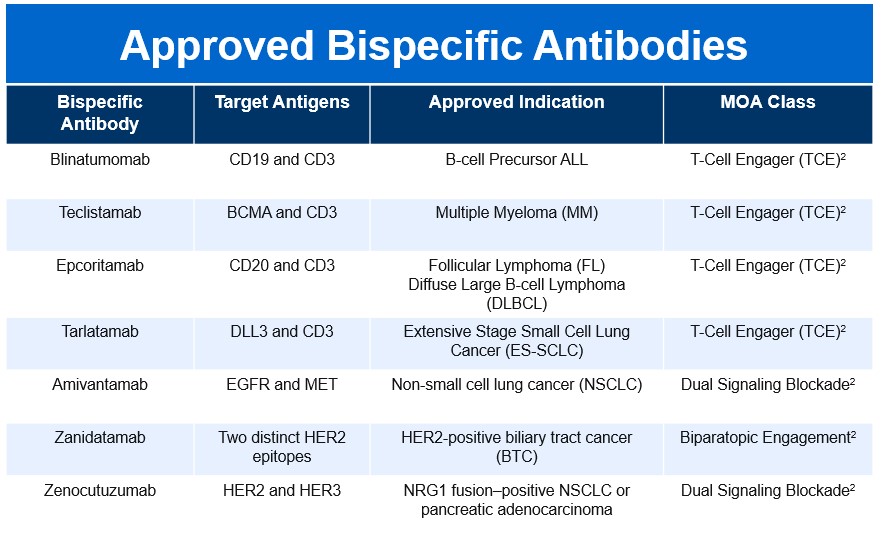
The BsAb landscape has rapidly expanded since the first approval of Blinatumomab in 2014, reflecting a growing therapeutic impact across multiple disease areas. As of late 2025, fifteen bispecific molecules have secured FDA approval, spanning both oncology and non-oncology indications. This surge underscores the versatility of BsAbs and their ability to address complex biological pathways through innovative mechanisms of action.

Approved-Bispecific-Antibodies

Limitations, Safety, and Risk Mitigation

  1. Manufacturing Challenges: The inherent complexity of BsAbs introduces challenges related to stability, manufacturability, and impurity control.1 The fusion of exogenous antigen-binding domains can decrease biophysical stability, and the complex assembly process frequently results in the formation of product-related impurities and mispaired species, which are difficult to remove during purification. These factors are not merely manufacturing hurdles; they directly influence the biological activity and, critically, the immunogenic potential of the final drug product.
  2. MOA-Specific Toxicity: The safety profile of BsAbs is highly dependent on their MOA
    • T-Cell Engager Toxicity: The highly potent, acute T-cell activation triggered by TCEs results in two major, distinct safety concerns. The first is Cytokine Release Syndrome (CRS), a serious acute toxicity caused by the mass release of systemic cytokines. CRS has been reported in up to 70% of patients receiving BsAbs, often necessitating hospitalization and precise management protocols. Severe cases (Grade ≥ 3) occur in 5–10% of patients.6 The second concern is Neurotoxicity (ICANS), which, while less frequent than CRS, affects 10–15% of patients and can range from mild confusion to cerebral edema.6 In addition, there is an Immune Regulation Paradox. Paradoxically, T-BsAb therapy can trigger the expansion and activation of inhibitory Regulatory T (Treg) cells in the tumor microenvironment, leading to the production of anti-inflammatory cytokines like IL-10. This critically inhibits the desired effector T-cell response, suggesting that combination strategies—such as transient Treg ablation—may be necessary to maximize efficacy.6
    • Pathway Blocker and Biparatopic Toxicity: These agents generally do not induce acute, systemic cytokine surges. Instead, their adverse event profiles reflect the targeted receptors. For example, the dual signaling blocker Amivantamab (targets EGFR/MET) exhibits EGFR-inhibition-related dermatologic toxicities, like paronychia, skin fissures, and pruritus, as well as infusion reactions. Dual checkpoint inhibitors can have classic IO toxicities. Biparatopic antibodies, like Zanidatamab, demonstrate a manageable profile but frequently cause gastrointestinal toxicities (such as diarrhea and nausea/vomiting) and infusion-related reactions (IRRs).9 Importantly, clinical data for Zanidatamab confirmed no reports of CRS.9
  3. Mitigating Immunogenicity Risk: The complex structures, engineered sequences, and immunostimulatory MOAs of oncology BsAbs contribute to an increased risk of immunogenicity compared to mAbs. Mitigation must begin at the engineering stage, utilizing in silico prediction and in vitro assays to guide the selection of low-risk antibody constructs through deimmunization and tolerization methods.

Future Directions

The BsAb pipeline remains robust, reflecting a continuous drive toward addressing current clinical limitations and expanding into novel biological territories. The future of BsAbs is characterized by a strategic shift toward overcoming the immunosuppressive tumor microenvironment (TME). Emerging candidates are now focused on targets that modulate the innate immune system and TME suppression, such as LILRB1/2 bispecific IgG1 antibodies for advanced solid tumors.10-11 Furthermore, BsAbs are expanding beyond simple blockade, with molecules like SAR446422 (CD28xOX40 bispecific) in trial for inflammatory indications, demonstrating the potential for BsAbs to achieve synergistic co-stimulatory agonism.10-11 The continuous innovation in structural design, focused now on minimizing impurity-driven immunogenicity and maximizing the therapeutic window, ensures that BsAbs are poised to become the standard for highly tailored, multifunctional therapeutic intervention across diverse and complex diseases. The future of BsAbs is very promising.10-11

References:

  1. Shan KS, Musleh Ud Din S, Dalal S, Gonzalez T, Dalal M, Ferraro P, Hussein A, Vulfovich M. Bispecific Antibodies in Solid Tumors: Advances and Challenges. International Journal of Molecular Sciences. 2025; 26(12):5838. https://doi.org/10.3390/ijms26125838.
  2. The Bispecific 2024 Landscape Review. Beacon Intelligence. 2024. https://beacon-intelligence.com/landscape-reviews/bispecific/. Accessed November 23, 2025.
  3. Ai Z, Wang B, Song Y, Cheng P, Liu X, Sun P. Prodrug-based bispecific antibodies for cancer therapy: advances and future directions. Front Immunol. 2025;16:1523693. Published 2025 Jan 22. doi:10.3389/fimmu.2025.1523693.
  4. Amash A, Volkers G, Farber P, et al. Developability considerations for bispecific and multispecific antibodies. MAbs. 2024;16(1):2394229. doi:10.1080/19420862.2024.2394229.
  5. Shui L, Wu D, Yang K, Sun C, Li Q, Yin R. Bispecific antibodies: unleashing a new era in oncology treatment. Mol Cancer. 2025;24(1):212. Published 2025 Aug 4. doi:10.1186/s12943-025-02390-y.
  6. Dewaele L, Fernandes RA. Bispecific T-cell engagers for the recruitment of T cells in solid tumors: a literature review. Immunother Adv. 2025;5(1):ltae005. Published 2025 Jan 27. doi:10.1093/immadv/ltae005.
  7. Kast F, Schwill M, Stüber JC, et al. Engineering an anti-HER2 biparatopic antibody with a multimodal mechanism of action. Nat Commun. 2021;12(1):3790. Published 2021 Jun 18. doi:10.1038/s41467-021-23948-6.
  8. Elimova E, Ajani J, Burris H, et al. Zanidatamab plus chemotherapy as first-line treatment for patients with HER2-positive advanced gastro-oesophageal adenocarcinoma: primary results of a multicentre, single-arm, phase 2 study. Lancet Oncol. 2025;26(7):847-859. doi:10.1016/S1470-2045(25)00287-6.
  9. Ziihera Safety Information. Ziihera HCP (Jazz Pharmaceuticals). https://www.ziiherahcp.com/safety. Accessed November 23, 2025.
  10. Wen J, Cui W, Yin X, et al. Application and future prospects of bispecific antibodies in the treatment of non-small cell lung cancer. Cancer Biol Med. 2025;22(4):348-375. doi:10.20892/j.issn.2095-3941.2024.0470.
  11. Engineering the Next Generation of Bispecific Antibodies. PEGS Europe 2024 Archive. https://www.pegsummiteurope.com/24/engineering-bispecifics. Accessed November 23, 2025

Educational Message: Precision Medicine in mCRC: Navigating Complexity in the Era of Targeted Therapy

Written by: David Cosgrove, MD
Sponsored by: Takeda

Treatment algorithms for patients with metastatic colorectal cancer (mCRC) have become increasingly complex in recent years, as new drug approvals have created additional therapeutic avenues for specific subsets of patients.1 Traditionally, mCRC patients who are fit enough for active therapy would undergo combination cytotoxic chemotherapy utilizing a fluoropyrimidine backbone, until disease progression or unacceptable toxicity. There were a limited number of effective agents available, and specific combinations were selected primarily based on patient comorbidity and toxicity risk, rather than on any tumor-specific factors. Predictably, within this model, response rates were modest and long-term outcomes remained grim.

Today, the majority of mCRC patients will still receive initial treatment with cytotoxic chemotherapy, but diagnostic testing is often incorporated to uncover actionable tumor-specific genomic and/or immune signatures, and these insights may be leveraged to guide the use of specific targeted therapies with improved patient outcomes.2,3,4 Information on tumor mismatch repair status (or microsatellite instability), specific mutations within KRAS/NRAS/BRAF, POLE/POLD-1, overexpression of HER2, and fusions within the NTRK gene all now contribute to treatment decisions at the time of diagnosis and at the time of disease progression where a treatment plan change is indicated. In addition to the recent approval of drugs to target these molecular signatures, an accompanying shift in drug formulation has impacted the mCRC treatment landscape.

Traditional mCRC anticancer agents were formulated for intravenous administration and delivered in an oncology office or hospital infusion suite. Dosing choices, supportive care medications, and treatment adjustments were typically decided by the treating oncologist, in conjunction with infusion nurses and the supporting clinical team; pharmacists played a role in dose confirmation, drug-drug interaction checks and admixture, but direct input beyond that was limited. Today, a majority of the new FDA-approved mCRC therapies are formulated for oral administration. Oral formulations free patients from being tethered to an infusion suite and alter the frequency and personnel involved in treatment touchpoints. The shift to oral formulations has expanded the role of pharmacy teams in patient education, dosing input, dispensing and toxicity assessment, while maintaining their role in drug safety.

Most oncology clinics have had to adapt their staffing and patient flow model to account for this new dynamic. Patient education is a key component to chemotherapy delivery – with traditional intravenous agents, infusion room nurses and oncology nurse educators typically took on this role, performing toxicity assessments and managing side effects chair-side. Traditionally, cytotoxic agents within the same drug class and mechanism of action often exhibited similar toxicity profiles, further simplifying toxicity risk assessments and corresponding patient education. With today’s newer, oral formulations, mechanisms of action and toxicity profiles are more varied – some retain cytotoxic effects, such as capecitabine or tipiracil/trifluridine, whereas others carry very specific toxicity profiles.

As patients may no longer receive treatment in an infusion suite, a significant portion of the responsibility for providing patient-level therapeutic education has been transferred to the pharmacist and pharmacy team. This educational role may be replicated through a series of subsequent treatments, as newer agents are typically delivered sequentially to these patients in later lines of therapy, depending on patient functional status, and suitability for ongoing treatment. Equally as important as pre-treatment education, on-therapy toxicity assessments and potential dose adjustments are now typically shared responsibilities between the treating physician and pharmacy team, and often incorporate patient reported outcomes (PROs) or electronic patient reported outcomes (ePROs)5, as the patient is taking these medications at home, and not under the direct supervision of an infusion nurse team as with the intravenous therapies.

Today’s mCRC treatment model requires close collaboration between the treating oncologist, who has typically developed a long-term therapeutic relationship with the patient and has knowledge of patient-specific factors that will influence treatment tolerance and potential side effects, and the pharmacy team. Lack of a robust communication system and/or improper delegation of tasks pose significant risks to the vulnerable mCRC patient population. To this end, many centers have developed Medically Integrated Pharmacies (MIP) for specialized oncology drugs, which provide direct oversight of quality and safety metrics, enhance adherence, reduce the risk of access delays and deliver appropriate patient-centered care. In our practice, we have seen countless examples of the MIP team lowering barriers to access, expediting delivery and intervening with dose adjustments or concomitant medication changes to ensure our mCRC patients glean as much benefit from their therapies as possible, while maintaining their desired quality of life in the face of a devastating illness.

As crucial as these aspects of care are for the treatment team, financial risk is a major concern for the mCRC patients themselves. Most of the newer therapies approved in the mCRC space in recent years are high-cost agents, and unlike intravenous agents, which were delivered in a medical facility and therefore covered under the medical benefit portion of a patient’s health insurance plan, oral formulation drugs fall under the pharmacy benefit. While we have seen fewer outright denials of coverage for clinically appropriate drugs, challenges remain such as prior authorization, onerous paperwork and especially patient co-payment requirements.

Unfortunately, a number of my patients have also faced barriers from their insurance-mandated, Limited Distribution Network (LDN), which incorporates an external Pharmacy Benefit Manager (PBM) and requires dispensing through a mail-order specialty pharmacy.6 The inability to communicate closely with LDNs and PBM-mandated third-party decision makers has proven challenging – without an on-site team to understand the specifics of a patient’s case, treating providers have limited ability to control dosing adjustments, maintain drug supply and limit care delays. Care delays pose very serious risks, especially in the later stages of mCRC during which dosing flexibility is critical and the majority of patients require dose holds or adjustments on a regular basis. While this issue remains to be solved, having an active MIP in a treatment center with dedicated staff to facilitate co-pay assistance and access to manufacturer- or foundation-level support has proven instrumental in many practices. This resource helps alleviate financial burden and ensures the patient is not forced to make therapy choices based on ability to pay when facing this illness.

In summary, therapeutic management of mCRC has become increasingly complex in recent years. The introduction of new therapeutic agents offers renewed hope for patients dealing with this devastating disease, while simultaneously requiring oncology practices to adjust treatment team infrastructure, and has shifted the onus of delivering patient education to the pharmacy team, who must work in close collaboration with the treating physician. Today’s shift to oral drug formulations introduces financial risks for patients, as at-home medications fall under a prescription drug benefit which may introduce additional barriers such as may PBM-mandated LDNs or specialty pharmacy requirements. The creation of MIPs has significantly enhanced provider communication, reduced barriers to access, expedited therapy delivery, and supported timely dose adjustments or medication changes to help mCRC patients gain the most benefit from treatment. MIPs have also been essential in building a broader administrative team focused on ensuring patients receive maximum benefit from breakthrough anticancer agents, while minimizing both physical and financial toxicity.

References:

  1. https://www.nccn.org/professionals/physician_gls/pdf/colon.pdf
  2. Yaeger R, Weiss J, Pelster M, et al. Adagrasib with or without Cetuximab in colorectal cancer with mutated KRAS G12C. N Engl J Med 2023;388:44-54
  3. Kopetz S, Yoshino T, Cutsem EV, et al. Encorafenib, cetuximab and chemotherapy in BRAF-mutant colorectal cancer: a randomized phase 3 trial. Nat Med 2025
  4. Overman MJ, Lonardi S, Wong K, et al. Durable clinical benefit with nivolumab plus ipilimumab in DNA mismatch repair-deficient/microsatellite instability-high metastatic colorectal cancer. J Clin Oncol 2018;36:773-779
  5. Basch E, Deal A, et al. Overall survival results of a trial assessing patient-reported outcomes for symptom monitoring during routine cancer treatment. JAMA 2017 July 11;318(2):197-198
  6. https://www.ncoda.org/oold/

Maintaining Physician Preference in CLL Care and Its Impact on Outcomes

Written by: M. Yair Levy, MD
Sponsored by: BeOne Medicines

Chronic lymphocytic leukemia (CLL) is a heterogeneous disease that has seen a remarkable treatment evolution over the past decade. Approximately one-third of CLL patients will not require treatment, with clinical observation remaining the standard for asymptomatic patients.1 For CLL patients requiring treatment, historical regimens have been limited to cytotoxic chemotherapy, administered alone or in combination with anti-CD20 immunotherapy.2 In 2016, two classes of targeted therapies entered the CLL marketplace. Most notably among these are inhibitors of the anti-apoptotic protein B-cell lymphoma 2 (Bcl2), and the B-cell proliferative regulator Bruton’s tyrosine kinase (BTK).2 Introduction of first-generation covalent BTK inhibitors (cBTKis) was followed by second-generation, as well as non-covalent BTKis.2 The rapid expansion of targeted therapies over the last decade has profoundly impacted the clinical management of CLL.

The multitude of therapeutic options in today’s CLL treatment landscape, which often includes several drugs in a single class, provides oncologists the unprecedented opportunity to weigh the entire clinical picture for each patient, and personalize their treatment strategy accordingly. Clinicians leverage decades of medical training to gauge pharmacokinetic and pharmacodynamic differences between therapies, weighing drug-specific efficacy, tolerability and safety for each patient. There is also concurrent influence from reputable medical bodies, such as NCCN guidelines and FDA approved treatments. Most importantly, every CLL patient and their disease are unique. It is imperative to protect physicians’ decision-making freedom to the degree that it permits individualized treatment approaches and the prescription of specific therapies, without undue external influence.

Step therapy is a growing utilization management strategy among health plan Pharmacy Benefit Managers (PBMs) that threatens physician autonomy. As its colloquial name “fail-first” implies, step therapy is a cost-saving approach that requires a patient to have tried, and failed, alternative drug(s), before the PBM will cover the originally prescribed drug.3 Step therapies transfer the onus of decision-making from the treating physician to a commercial entity, robbing the clinician of their ability to personalize care and maximize patient outcomes. This leads to worsening provider burnout, depersonalization, and exacerbates oncology physician shortages. Step therapies also place additional burden on clinical support staff and negatively impact CLL patient outcomes.

PBM-mandated BTKi coverage is a common scenario that exemplifies how step therapy negatively impacts CLL patient outcomes. In my clinical experience, PBMs have required frontline use of the first-generation BTKi ibrutinib. Ibrutinib is an efficacious drug that has revolutionized CLL treatment, but it is poorly tolerated, as compared with 2nd generation BTKis, which directly contributes to decreased treatment compliance and impacts patient outcomes. Ibrutinib also carries the risk of adverse cardiologic events, including atrial arrythmias, ventricular arrhythmias and sudden death.4 As a provider who has lost a patient due to this adverse event, it’s imperative that safety risks of specific drugs are weighed by an experienced clinician on a case-to-case basis. Furthermore, there is an urgent need for step therapy reform that considers clinical data in determining drug coverage. In the case of BTKi coverage, there is clinical evidence demonstrating a lower risk of adverse cardiologic events with use of second-generation BTKi agents.4 As cancer patient advocates, oncologists must continue to push back against step therapies that infringe on patient safety and well-being, and may adversely affect outcomes.

As oncology drugs continue to enter the ever-changing healthcare marketplace, it’s increasingly critical that physicians communicate the importance of preserving autonomy to PBMs and other decision-making parties. There is potential for other drug pricing provisions, such as those included in the 2022 Inflation Reduction Act (IRA),5 to impact CLL patient outcomes in a manner similar to that which is seen with PBM-mediated step therapy. Under the IRA, the government may select certain medications for which it will cap the cost of for Medicare recipients.5 While designed to reduce patient financial burden, provisions of the IRA, such as the ability to target costs of small molecules (like BTKis) years before other drug types, may have harmful effects on oncology treatment.5 This risks physicians being forced to utilize certain medications for which cost setting has been established, similar to step therapy, and has already been shown to disincentivize the development of small molecule drugs since the IRA was enacted.5 Given that CLL primarily affects elderly populations,2 drug pricing provisions affecting Medicare could impact CLL prescribing patterns, and there is precedent to surmise that commercial payers may adopt similar strategies of government-sponsored programs.

In diseases like CLL where therapeutic nuances matter, restricting access to preferred agents can have profound consequences. While the extra hours spent justifying what seems like every clinical decision, completing paperwork, appealing denials, and engaging in peer-to-peer calls may feel like “death by a thousand paper cuts”, oncologists have the responsibility as patient advocates to do no harm. We must continue to advocate for policies that prioritize patient outcomes over cost-driven algorithms, and ensure that clinical decision-making remains in the hands of those best equipped to make it: the treating physicians.

References:

  1. Shadman M. Diagnosis and Treatment of Chronic Lymphocytic Leukemia: A Review. JAMA. 2023;329(11):918-932. doi:10.1001/jama.2023.1946.
  2. Sekeres S, Lamkin EN, Bravo E Jr, Cool A, Taylor J. Resistance Mutations in CLL: Genetic Mechanisms Shaping the Future of Targeted Therapy. Genes (Basel). 2025;16(9):1064. Published 2025 Sep 10. doi:10.3390/genes16091064.
  3. Royce TJ, Schenkel C, Kirkwood K, Levit L, Levit K, Kircher S. Impact of Pharmacy Benefit Managers on Oncology Practices and Patients. JCO Oncol Pract. 2020;16(5):276-284. doi:10.1200/JOP.19.00606.
  4. Moslehi JJ, Furman RR, Tam CS, et al. Cardiovascular events reported in patients with B-cell malignancies treated with zanubrutinib. Blood Adv. 2024;8(10):2478-2490. doi:10.1182/bloodadvances.2023011641.
  5. The Inflation Reduction Act and Medicare Drug Price “Negotiation”. Pharmaceutical Research and Manufacturers of America (PhRMA) website. https://phrma.org/policy-issues/government-price-setting/inflation-reduction-act. Accessed October 1, 2025.

OPDIVO® (nivolumab) + YERVOY® (ipilimumab) in the first-line treatment of unresectable or metastatic HCC1-5

Treating 1L unresectable or metastatic HCC? 3-year data may help you reassess your approach
Explore this 1L dual immunotherapy option for eligible patients

Expert opinion: Aiwu Ruth He, MD, PhD*
*Dr Aiwu Ruth He, MD, PhD, is a paid consultant of Bristol Myers Squibb (BMS) who was compensated by BMS for her contributions to this article.
Content sponsored by Bristol Myers Squibb

Unmet need in unresectable or metastatic HCC
Hepatocellular carcinoma (HCC) accounts for 75–85% of primary liver cancer cases and unresectable or metastatic HCC (uHCC) is associated with poor prognosis4,6. Thus, additional 1L treatments that prolong survival are needed. Approved 1L options, e.g. I-O + VEGFi and a dual I-O option, were evaluated against sorafenib alone, while OPDIVO® + YERVOY® was evaluated against investigator’s choice of lenvatinib or sorafenib, offering an alternative 1L immunotherapy option for uHCC1-5.

OPDIVO + YERVOY are associated with the following Warnings and Precautions2: severe and fatal immune-mediated adverse reactions including pneumonitis, colitis, hepatitis and hepatotoxicity, endocrinopathies, nephritis with renal dysfunction, dermatologic adverse reactions, other immune-mediated adverse reactions; infusion-related reactions; complications of allogeneic hematopoietic stem cell transplantation (HSCT); embryo-fetal toxicity; and increased mortality in patients with multiple myeloma when OPDIVO is added to a thalidomide analogue and dexamethasone, which is not recommended outside of controlled clinical trials.

Please see additional Important Safety Information for OPDIVO and YERVOY below, and U.S. Full Prescribing Information for OPDIVO and YERVOY.

OPDIVO + YERVOY in 1L treatment of unresectable or metastatic HCC
OPDIVO + YERVOY, a dual immune-checkpoint inhibitor combination studied in the global, randomized, phase 3 Checkmate 9DW trial, is the only FDA-approved treatment to show positive results* versus investigator’s choice of lenvatinib or sorafenib, in the 1L treatment of adult patients with uHCC1-5. Checkmate 9DW enrolled 668 patients with uHCC who were systemic-therapy–naïve, with at least 1 measurable lesion, Child-Pugh score 5 or 6, ECOG PS 0 or 1, and no main portal vein invasion (Vp4)1-3. An esophagogastroduodenoscopy (EGD) was not required2. Patients were randomized 1:1 to receive either OPDIVO (1 mg/kg) + YERVOY (3 mg/kg) q3w for up to 4 cycles followed by OPDIVO (480 mg IV) q4w or investigator’s choice of lenvatinib or sorafenib in the comparator arm1,2,5. YERVOY 3 mg/kg dosing in Checkmate 9DW was supported by Checkmate 0407.

HCC-Only-FDA-Approved-Treatment-Positive_Results

Efficacy and safety data for OPDIVO + YERVOY
Median overall survival (mOS), the primary endpoint, was 23.7 months (95% CI: 18.8, 29.4) with OPDIVO + YERVOY versus 20.6 months (95% CI: 17.5, 22.5) with the comparator arm, with a hazard ratio of 0.79 (95% CI: 0.65, 0.96; P=0.018)1,2. Notably, 38% of patients were alive at 3 years with OPDIVO + YERVOY, compared to 24% with the comparator arm1-3,5. According to Dr. He, “The Checkmate 9DW design was unique because one of the comparator arm treatments was investigator’s choice of lenvatinib—a modern TKI, received by 85% of patients—making these results particularly meaningful.”

OPDIVO + YERVOY achieved a deeper overall response rate (ORR), a secondary endpoint, of 36%, versus 13% with the comparator arm (P<0.0001)1-3,5. “The ORR being higher than the comparator arm is great. When my patients are symptomatic with a high tumor burden, I choose treatments with a higher response rate than their comparator arms to ease their symptoms and potentially reduce liver stress. Furthermore, an exploratory analysis found median time to response was 2.2 months,” said Dr. He.

Responses lasted over twice as long with the median duration of response (mDOR) at 30.4 months (95% CI: 21.2, NR) versus 12.9 months (95% CI: 10.2, 31.2) with the comparator arm1-3,5. Dr. He shared, “The DOR is clinically meaningful. Some of my Checkmate 9DW patients are now off treatment after 2 years, with ongoing disease control.”

Durable-Survival-Durable-Responses-Longer-Responses

The safety profile of OPDIVO + YERVOY is well established2,5. Dr. He added, “No new IMARs were observed in Checkmate 9DW. I encourage patients to report side effects early, and I manage IMARs per established protocols including treatment holds and steroid use.”

Opdivo-Yervoy-Safety

Summary and conclusions
OPDIVO + YERVOY, a differentiated 1L option for treatment of uHCC, achieved durable survival, deeper and longer responses with a well-established safety profile. “For OPDIVO + YERVOY, I choose patients who are relatively robust, socially supported, and report side effects promptly. I’m excited about this 1L treatment and its demonstrated durable survival benefits,” noted Dr. He.

INDICATIONS

OPDIVO, in combination with YERVOY, is indicated for the first-line treatment of adult patients with unresectable or metastatic hepatocellular carcinoma (HCC).

IMPORTANT SAFETY INFORMATION

Severe and Fatal Immune-Mediated Adverse Reactions

  • Immune-mediated adverse reactions listed herein may not include all possible severe and fatal immune-mediated adverse reactions.
  • Immune-mediated adverse reactions, which may be severe or fatal, can occur in any organ system or tissue. While immune-mediated adverse reactions usually manifest during treatment, they can also occur after discontinuation of OPDIVO or YERVOY. Early identification and management are essential to ensure safe use of OPDIVO and YERVOY. Monitor for signs and symptoms that may be clinical manifestations of underlying immune-mediated adverse reactions. Evaluate clinical chemistries including liver enzymes, creatinine, adrenocorticotropic hormone (ACTH) level, and thyroid function at baseline and periodically during treatment with OPDIVO and before each dose of YERVOY. In cases of suspected immune-mediated adverse reactions, initiate appropriate workup to exclude alternative etiologies, including infection. Institute medical management promptly, including specialty consultation as appropriate.
  • Withhold or permanently discontinue OPDIVO and YERVOY depending on severity (please see section 2 Dosage and Administration in the accompanying Full Prescribing Information). In general, if OPDIVO or YERVOY interruption or discontinuation is required, administer systemic corticosteroid therapy (1 to 2 mg/kg/day prednisone or equivalent) until improvement to Grade 1 or less. Upon improvement to Grade 1 or less, initiate corticosteroid taper and continue to taper over at least 1 month. Consider administration of other systemic immunosuppressants in patients whose immune-mediated adverse reactions are not controlled with corticosteroid therapy. Toxicity management guidelines for adverse reactions that do not necessarily require systemic steroids (e.g., endocrinopathies and dermatologic reactions) are discussed below.

Immune-Mediated Pneumonitis

  • OPDIVO and YERVOY can cause immune-mediated pneumonitis. The incidence of pneumonitis is higher in patients who have received prior thoracic radiation. In patients receiving OPDIVO 1 mg/kg with YERVOY 3 mg/kg every 3 weeks, immune-mediated pneumonitis occurred in 7% (31/456) of patients, including Grade 4 (0.2%), Grade 3 (2.0%), and Grade 2 (4.4%).

Immune-Mediated Colitis

  • OPDIVO and YERVOY can cause immune-mediated colitis, which may be fatal. A common symptom included in the definition of colitis was diarrhea. Cytomegalovirus (CMV) infection/reactivation has been reported in patients with corticosteroid-refractory immune-mediated colitis. In cases of corticosteroid-refractory colitis, consider repeating infectious workup to exclude alternative etiologies. In patients receiving OPDIVO 1 mg/kg with YERVOY 3 mg/kg every 3 weeks, immune-mediated colitis occurred in 25% (115/456) of patients, including Grade 4 (0.4%), Grade 3 (14%) and Grade 2 (8%).

Immune-Mediated Hepatitis and Hepatotoxicity

  • OPDIVO and YERVOY can cause immune-mediated hepatitis. In patients receiving OPDIVO 1 mg/kg with YERVOY 3 mg/kg every 3 weeks, immune-mediated hepatitis occurred in 15% (70/456) of patients, including Grade 4 (2.4%), Grade 3 (11%), and Grade 2 (1.8%).

Immune-Mediated Endocrinopathies

  • OPDIVO and YERVOY can cause primary or secondary adrenal insufficiency, immune-mediated hypophysitis, immune-mediated thyroid disorders, and Type 1 diabetes mellitus, which can present with diabetic ketoacidosis. Withhold OPDIVO and YERVOY depending on severity (please see section 2 Dosage and Administration in the accompanying Full Prescribing Information). For Grade 2 or higher adrenal insufficiency, initiate symptomatic treatment, including hormone replacement as clinically indicated. Hypophysitis can present with acute symptoms associated with mass effect such as headache, photophobia, or visual field defects. Hypophysitis can cause hypopituitarism; initiate hormone replacement as clinically indicated. Thyroiditis can present with or without endocrinopathy. Hypothyroidism can follow hyperthyroidism; initiate hormone replacement or medical management as clinically indicated. Monitor patients for hyperglycemia or other signs and symptoms of diabetes; initiate treatment with insulin as clinically indicated.
  • In patients receiving OPDIVO 1 mg/kg with YERVOY 3 mg/kg every 3 weeks, adrenal insufficiency occurred in 8% (35/456) of patients, including Grade 4 (0.2%), Grade 3 (2.4%), and Grade 2 (4.2%).
  • In patients receiving OPDIVO 1 mg/kg with YERVOY 3 mg/kg every 3 weeks, hypophysitis occurred in 9% (42/456) of patients, including Grade 3 (2.4%) and Grade 2 (6%).
  • In patients receiving OPDIVO 1 mg/kg with YERVOY 3 mg/kg every 3 weeks, hyperthyroidism occurred in 9% (42/456) of patients, including Grade 3 (0.9%) and Grade 2 (4.2%).
  • In patients receiving OPDIVO 1 mg/kg with YERVOY 3 mg/kg every 3 weeks, hypothyroidism occurred in 20% (91/456) of patients, including Grade 3 (0.4%) and Grade 2 (11%).

Immune-Mediated Nephritis with Renal Dysfunction

  • OPDIVO and YERVOY can cause immune-mediated nephritis.

Immune-Mediated Dermatologic Adverse Reactions

  • OPDIVO can cause immune-mediated rash or dermatitis. Exfoliative dermatitis, including Stevens-Johnson syndrome (SJS), toxic epidermal necrolysis (TEN), and drug rash with eosinophilia and systemic symptoms (DRESS) has occurred with PD-1/PD-L1 blocking antibodies. Topical emollients and/or topical corticosteroids may be adequate to treat mild to moderate non-exfoliative rashes.
  • YERVOY can cause immune-mediated rash or dermatitis, including bullous and exfoliative dermatitis, SJS, TEN, and DRESS. Topical emollients and/or topical corticosteroids may be adequate to treat mild to moderate non-bullous/exfoliative rashes.
  • Withhold or permanently discontinue OPDIVO and YERVOY depending on severity (please see section 2 Dosage and Administration in the accompanying Full Prescribing Information).
  • In patients receiving OPDIVO 1 mg/kg with YERVOY 3 mg/kg every 3 weeks, immune-mediated rash occurred in 28% (127/456) of patients, including Grade 3 (4.8%) and Grade 2 (10%).

Other Immune-Mediated Adverse Reactions

  • The following clinically significant immune-mediated adverse reactions occurred at an incidence of <1% (unless otherwise noted) in patients who received OPDIVO monotherapy or OPDIVO in combination with YERVOY or were reported with the use of other PD-1/PD-L1 blocking antibodies. Severe or fatal cases have been reported for some of these adverse reactions: cardiac/vascular: myocarditis, pericarditis, vasculitis; nervous system: meningitis, encephalitis, myelitis and demyelination, myasthenic syndrome/myasthenia gravis (including exacerbation), Guillain-Barré syndrome, nerve paresis, autoimmune neuropathy; ocular: uveitis, iritis, and other ocular inflammatory toxicities can occur; gastrointestinal: pancreatitis to include increases in serum amylase and lipase levels, gastritis, duodenitis; musculoskeletal and connective tissue: myositis/polymyositis, rhabdomyolysis, and associated sequelae including renal failure, arthritis, polymyalgia rheumatica; endocrine: hypoparathyroidism; other (hematologic/immune): hemolytic anemia, aplastic anemia, hemophagocytic lymphohistiocytosis (HLH), systemic inflammatory response syndrome, histiocytic necrotizing lymphadenitis (Kikuchi lymphadenitis), sarcoidosis, immune thrombocytopenic purpura, solid organ transplant rejection, other transplant (including corneal graft) rejection.
  • In addition to the immune-mediated adverse reactions listed above, across clinical trials of YERVOY monotherapy or in combination with OPDIVO, the following clinically significant immune-mediated adverse reactions, some with fatal outcome, occurred in <1% of patients unless otherwise specified: nervous system: autoimmune neuropathy (2%), myasthenic syndrome/myasthenia gravis, motor dysfunction; cardiovascular: angiopathy, temporal arteritis; ocular: blepharitis, episcleritis, orbital myositis, scleritis; gastrointestinal: pancreatitis (1.3%); other (hematologic/immune): conjunctivitis, cytopenias (2.5%), eosinophilia (2.1%), erythema multiforme, hypersensitivity vasculitis, neurosensory hypoacusis, psoriasis.
  • Some ocular IMAR cases can be associated with retinal detachment. Various grades of visual impairment, including blindness, can occur. If uveitis occurs in combination with other immune-mediated adverse reactions, consider a Vogt-Koyanagi-Harada–like syndrome, which has been observed in patients receiving OPDIVO and YERVOY, as this may require treatment with systemic corticosteroids to reduce the risk of permanent vision loss.

Infusion-Related Reactions

  • OPDIVO and YERVOY can cause severe infusion-related reactions. Discontinue OPDIVO and YERVOY in patients with severe (Grade 3) or life-threatening (Grade 4) infusion-related reactions. Interrupt or slow the rate of infusion in patients with mild (Grade 1) or moderate (Grade 2) infusion-related reactions. In HCC patients receiving OPDIVO 1 mg/kg with YERVOY 3 mg/kg every 3 weeks, infusion-related reactions occurred in 8% (4/49) of patients.

Complications of Allogeneic Hematopoietic Stem Cell Transplantation

  • Fatal and other serious complications can occur in patients who receive allogeneic hematopoietic stem cell transplantation (HSCT) before or after being treated with OPDIVO or YERVOY. Transplant-related complications include hyperacute graft-versus-host-disease (GVHD), acute GVHD, chronic GVHD, hepatic veno-occlusive disease (VOD) after reduced intensity conditioning, and steroid-requiring febrile syndrome (without an identified infectious cause). These complications may occur despite intervening therapy between OPDIVO or YERVOY and allogeneic HSCT.
  • Follow patients closely for evidence of transplant-related complications and intervene promptly. Consider the benefit versus risks of treatment with OPDIVO and YERVOY prior to or after an allogeneic HSCT.

Embryo-Fetal Toxicity

  • Based on its mechanism of action and findings from animal studies, OPDIVO and YERVOY can cause fetal harm when administered to a pregnant woman. The effects of YERVOY are likely to be greater during the second and third trimesters of pregnancy. Advise pregnant women of the potential risk to a fetus. Advise females of reproductive potential to use effective contraception during treatment with OPDIVO and YERVOY and for at least 5 months after the last dose.

Increased Mortality in Patients with Multiple Myeloma when OPDIVO is Added to a Thalidomide Analogue and Dexamethasone

  • In randomized clinical trials in patients with multiple myeloma, the addition of OPDIVO to a thalidomide analogue plus dexamethasone resulted in increased mortality. Treatment of patients with multiple myeloma with a PD-1 or PD-L1 blocking antibody in combination with a thalidomide analogue plus dexamethasone is not recommended outside of controlled clinical trials.

Lactation

  • There are no data on the presence of OPDIVO or YERVOY in human milk, the effects on the breastfed child, or the effects on milk production. Because of the potential for serious adverse reactions in breastfed children, advise women not to breastfeed during treatment and for 5 months after the last dose.

Serious Adverse Reactions

  • In Checkmate 9DW, serious adverse reactions occurred in 53% of patients receiving OPDIVO with YERVOY (n=332). The most frequent non liver-related serious adverse reactions reported in ≥2% of patients who received OPDIVO with YERVOY were diarrhea/colitis (4.5%), gastrointestinal hemorrhage (3%), and rash (2.4%). Liver-related serious adverse reactions occurred in 17% of patients receiving OPDIVO with YERVOY, including Grade 3-4 events in 16% of patients. The most frequently reported all grade liver-related serious adverse reactions occurring in ≥1% of patients who received OPDIVO with YERVOY were immune-mediated hepatitis (3%), increased AST/ALT (3%), hepatic failure (2.4%), ascites (2.4%), and hepatotoxicity (1.2%). Fatal adverse reactions occurred in 12 (3.6%) patients who received OPDIVO with YERVOY; these included 4 (1.2%) patients who died due to immune-mediated or autoimmune hepatitis and 4 (1.2%) patients who died of hepatic failure.

Common Adverse Reactions

  • In Checkmate 9DW, the most common adverse reactions (≥20%) in patients receiving OPDIVO with YERVOY (n=332) were rash (36%), pruritus (34%), fatigue (33%), and diarrhea (25%).

Clinical Trials and Patient Populations

  • Checkmate 9DW – hepatocellular carcinoma, in combination with YERVOY.


By unsubscribing from OncoPrescribe.com below, you are not opting out of receiving email marketing communications from Bristol-Myers Squibb Company.

References

  1. Kudo M et al. Oral presentation at ASCO-GI 2025. Abstract 520.
  2. OPDIVO [package insert]. Princeton, NJ; Bristol-Myers Squibb Company.
  3. Galle PR et al. Oral presentation at ASCO 2024. Abstract LBA4008.
  4. American Cancer Society. https://www.cancer.org/cancer/liver-cancer/detection-diagnosis-staging/survival-rates.html. Accessed July 2025.
  5. YERVOY [package insert]. Princeton, NJ; Bristol-Myers Squibb Company.
  6. Yau T et al. 2025;405(10492):1851–1864.
  7. El-Khoueiry AB et al. Lancet. 2017;389(10088):2492–2502.
  8. Data on file. BMS-REF-NIVO-0335. Princeton, NJ: Bristol-Myers Squibb Company; 2025.
  9. Data on file. BMS-REF-NIVO-0326. Princeton, NJ: Bristol-Myers Squibb Company; 2025.

© 2025 Bristol-Myers Squibb Company. OPDIVO® and YERVOY® are registered trademarks of Bristol-Myers Squibb Company.

7356-US-2500331   08/2025


OPDIVO QvantigTM (nivolumab + hyaluronidase-nvhy) is delivered via subcutaneous injection, streamlining administration for eligible patients1*

*3-5–minute vs 30-minute infusion of IV nivolumab. This does not account for all aspects of treatment. Actual clinic time may vary. 1,2

 Expert opinion: Saby George, MD, FACP
Dr Saby George, MD, is a paid consultant of Bristol Myers Squibb (BMS) who was compensated by BMS for his contributions to this article.

Content sponsored by Bristol Myers Squibb

Subcutaneous administration overview

While immune checkpoint inhibitors have emerged as key treatment options for certain types of cancer, they are primarily delivered through intravenous (IV) administration,3 creating a need for alternative routes of administration.4-5 A subcutaneous (SC) injection may reduce the time preparing and administering treatment compared to IV delivery, offer practice flexibility that may free up infusion chairs, and deliver treatment faster.3,5Infusion centers are overwhelmed. Infusion chairs may open up if we transition to approved SC options,” remarked Dr George.

Evaluation of comparable PK, efficacy, and safety of SC OPDIVO Qvantig with IV nivolumab

OPDIVO Qvantig is formulated with hyaluronidase to increase the dispersion and absorption of SC nivolumab.1 CheckMate 67T, a randomized, open-label, phase 3 noninferiority trial, was designed to compare the PK, efficacy, and safety of OPDIVO Qvantig (delivered as a SC injection) with IV nivolumab.1,4

 OPDIVO QVANTIG, as monotherapy, is indicated for the first-line treatment of adult patients with intermediate- or poor-risk advanced renal cell carcinoma (RCC), following treatment with intravenous nivolumab and ipilimumab combination therapy. OPDIVO QVANTIG is not indicated in combination with ipilimumab for the treatment of renal cell carcinoma. Please see additional 16 indications below.

OPDIVO QVANTIG is associated with the following Warnings and Precautions: severe and fatal immune-mediated adverse reactions including pneumonitis, colitis, hepatitis and hepatotoxicity, endocrinopathies, nephritis with renal dysfunction, dermatologic adverse reactions, other immune-mediated adverse reactions; complications of allogeneic hematopoietic stem cell transplantation (HSCT); embryo-fetal toxicity; and increased mortality in patients with multiple myeloma when OPDIVO QVANTIG is added to a thalidomide analogue and dexamethasone, which is not recommended outside of controlled clinical trials.

Please see Important Safety Information for OPDIVO QVANTIG below and US Full Prescribing Information for OPDIVO QVANTIG.

CheckMate 67T was a phase 3, randomized (1:1), open-label, noninferiority trial evaluating OPDIVO Qvantig (1,200 mg of nivolumab and 20,000 units of hyaluronidase) compared to intravenous nivolumab, in adult patients with advanced or metastatic clear-cell renal cell carcinoma (ccRCC) who received prior systemic therapy.1,4 Patients were stratified by weight (<80 kg versus ≥80 kg) and International Metastatic RCC Database Consortium (IMDC) risk score (favorable vs intermediate vs poor risk).1 A total of 495 patients were randomized to receive either OPDIVO Qvantig every 4 weeks subcutaneously (n=248) or nivolumab 3 mg/kg every 2 weeks intravenously (n=247).1,4 The co-primary endpoints were time-averaged serum concentration over 28 days (Cavgd28) and minimum serum concentration at steady state (Cminss).4 The key powered secondary endpoint was overall response rate, as assessed by blinded independent central review. The minimum follow-up time was 8 months.4

Pharmacokinetic, efficacy, and safety results

CheckMate 67T demonstrated that the PK of OPDIVO Qvantig was noninferior to that of intravenously administered nivolumab.1,4*


OPDIVO Qvantig resulted in a safety profile comparable with IV nivolumab.1 Dr George noted, “Safety was similar between administration methods. Rates of adverse reactions were similar for IV and SC nivolumab administration.6 Please see safety table below for more information.

Summary and conclusions

OPDIVO Qvantig resulted in comparable PK, efficacy, and safety to IV nivolumab and may be the right option for your eligible patients.1 This 3-5 minute SC injection option may reduce the steps required for preparation and time needed for administration compared to IV nivolumab.1,2* There is no need for IV preparation, dilution, weight-based dose calculations, or port access with OPDIVO Qvantig.1 According to Dr George, “For my appropriate patients, it gives me flexibility. It may save administration time.* For eligible patients, it’s great to have this subcutaneous treatment option.

*3-5–minute vs 30-minute infusion of IV nivolumab. This does not account for all aspects of treatment. Actual clinic time may vary.1,2

INDICATIONS

OPDIVO QVANTIG™ (nivolumab and hyaluronidase-nvhy), as monotherapy, is indicated for the first-line treatment of adult patients with intermediate- or poor-risk advanced renal cell carcinoma (RCC), following treatment with intravenous nivolumab and ipilimumab combination therapy.
Limitations of Use: OPDIVO QVANTIG is not indicated in combination with ipilimumab for the treatment of renal cell carcinoma.

OPDIVO QVANTIG™ (nivolumab and hyaluronidase-nvhy), in combination with cabozantinib, is indicated for the first-line treatment of adult patients with advanced renal cell carcinoma (RCC).

OPDIVO QVANTIG™ (nivolumab and hyaluronidase-nvhy), as monotherapy, is indicated for the treatment of adult patients with advanced renal cell carcinoma (RCC) who have received prior anti-angiogenic therapy.

OPDIVO QVANTIG™ (nivolumab and hyaluronidase-nvhy), as monotherapy, is indicated for the treatment of adult patients with unresectable or metastatic melanoma.

OPDIVO QVANTIG™ (nivolumab and hyaluronidase-nvhy), as monotherapy, is indicated for the treatment of adult patients with unresectable or metastatic melanoma following treatment with intravenous nivolumab and ipilimumab combination therapy.
Limitations of Use: OPDIVO QVANTIG is not indicated in combination with ipilimumab for treatment of unresectable or metastatic melanoma.

OPDIVO QVANTIG™ (nivolumab and hyaluronidase-nvhy), as monotherapy, is indicated for the adjuvant treatment of adult patients with completely resected Stage IIB, Stage IIC, Stage III, or Stage IV melanoma.

OPDIVO QVANTIG™ (nivolumab and hyaluronidase-nvhy), in combination with platinum-doublet chemotherapy, is indicated as neoadjuvant treatment of adult patients with resectable (tumors ≥4 cm or node positive) non-small cell lung cancer (NSCLC).

OPDIVO QVANTIG™ (nivolumab and hyaluronidase-nvhy), in combination with platinum-doublet chemotherapy, is indicated for the neoadjuvant treatment of adult patients with resectable (tumors ≥4 cm or node positive) non-small cell lung cancer (NSCLC) and no known epidermal growth factor receptor (EGFR) mutations or anaplastic lymphoma kinase (ALK) rearrangements, followed by OPDIVO QVANTIG as monotherapy in the adjuvant setting after surgical resection.

OPDIVO QVANTIG™ (nivolumab and hyaluronidase-nvhy), as monotherapy, is indicated for the treatment of adult patients with metastatic non-small cell lung cancer (NSCLC) with progression on or after platinum-based chemotherapy. Patients with EGFR or ALK genomic tumor aberrations should have disease progression on FDA-approved therapy for these aberrations prior to receiving OPDIVO QVANTIG.
Limitations of Use: OPDIVO QVANTIG is not indicated in combination with ipilimumab for the treatment of metastatic NSCLC.

OPDIVO QVANTIG™ (nivolumab and hyaluronidase-nvhy), as monotherapy, is indicated for the treatment of adult patients with recurrent or metastatic squamous cell carcinoma of the head and neck (SCCHN) with disease progression on or after platinum-based therapy.

OPDIVO QVANTIG™ (nivolumab and hyaluronidase-nvhy), as monotherapy, is indicated for the adjuvant treatment of adult patients with urothelial carcinoma (UC) who are at high risk of recurrence after undergoing radical resection of UC.

OPDIVO QVANTIG™ (nivolumab and hyaluronidase-nvhy), in combination with cisplatin and gemcitabine, is indicated for the first-line treatment of adult patients with unresectable or metastatic urothelial carcinoma (UC).

OPDIVO QVANTIG™ (nivolumab and hyaluronidase-nvhy), as monotherapy, is indicated for the treatment of adult patients with locally advanced or metastatic urothelial carcinoma (UC) who have disease progression during or following platinum-containing chemotherapy or have disease progression within 12 months of neoadjuvant or adjuvant treatment with platinum-containing chemotherapy.

OPDIVO QVANTIG™ (nivolumab and hyaluronidase-nvhy), as monotherapy, is indicated for the adjuvant treatment of completely resected esophageal or gastroesophageal junction cancer with residual pathologic disease in adult patients who have received neoadjuvant
chemoradiotherapy (CRT).

OPDIVO QVANTIG™ (nivolumab and hyaluronidase-nvhy), in combination with fluoropyrimidine- and platinum-containing chemotherapy, is indicated for the first-line treatment of adult patients with unresectable advanced or metastatic esophageal squamous cell carcinoma (ESCC) whose tumors express PD-L1 (≥1%).
Limitations of Use: OPDIVO QVANTIG is not indicated in combination with ipilimumab for the treatment of patients with unresectable advanced or metastatic ESCC.

OPDIVO QVANTIG™ (nivolumab and hyaluronidase-nvhy), as monotherapy, is indicated for the treatment of adult patients with unresectable advanced, recurrent or metastatic esophageal squamous cell carcinoma (ESCC) after prior fluoropyrimidine-and platinum-based chemotherapy.

OPDIVO QVANTIG™ (nivolumab and hyaluronidase-nvhy), in combination with fluoropyrimidine- and platinum-containing chemotherapy, is indicated for the treatment of adult patients with advanced or metastatic gastric cancer, gastroesophageal junction cancer, and esophageal adenocarcinoma whose tumors express PD-L1 (≥1%).

IMPORTANT SAFETY INFORMATION 

Severe and Fatal Immune-Mediated Adverse Reactions

  • Immune-mediated adverse reactions, which may be severe or fatal, can occur in any organ system or tissue. While immune-mediated adverse reactions usually manifest during treatment, they can also occur after discontinuation of OPDIVO QVANTIG. Early identification and management are essential to ensure safe use of OPDIVO QVANTIG. Monitor for signs and symptoms that may be clinical manifestations of underlying immune-mediated adverse reactions. Evaluate clinical chemistries including liver enzymes, creatinine, and thyroid function at baseline and periodically during treatment. In cases of suspected immune-mediated adverse reactions, initiate appropriate workup to exclude alternative etiologies, including infection. Institute medical management promptly, including specialty consultation as appropriate.
  • Withhold or permanently discontinue OPDIVO QVANTIG depending on severity (please see Section 2 Dosage and Administration in the accompanying Full Prescribing Information). In general, if OPDIVO QVANTIG interruption or discontinuation is required, administer systemic corticosteroid therapy (1 to 2 mg/kg/day prednisone or equivalent) until improvement to Grade 1 or less. Upon improvement to Grade 1 or less, initiate corticosteroid taper and continue to taper over for at least 1 month. Consider administration of other systemic immunosuppressants in patients whose immune-mediated adverse reactions are not controlled with corticosteroid therapy.
  • Toxicity management guidelines for adverse reactions that do not necessarily require systemic steroids (e.g., endocrinopathies and dermatologic reactions) are discussed below.

Immune-Mediated Pneumonitis

  • OPDIVO QVANTIG can cause immune-mediated pneumonitis. The incidence of pneumonitis is higher in patients who have received prior thoracic radiation.
  • Immune-mediated pneumonitis occurred in 2.8% (7/247) of patients receiving OPDIVO QVANTIG, including Grade 3 (0.8%) and Grade 2 (2.0%) adverse reactions.

Immune-Mediated Colitis

  • OPDIVO QVANTIG can cause immune-mediated colitis. A common symptom included in the definition of colitis was diarrhea. Cytomegalovirus (CMV) infection/reactivation has been reported in patients with corticosteroid-refractory immune-mediated colitis. In cases of corticosteroid-refractory colitis, consider repeating infectious workup to exclude alternative etiologies.
  • Immune-mediated colitis occurred in 2.8% (7/247) of patients receiving OPDIVO QVANTIG, including Grade 3 (0.4%) and Grade 2 (2.4%) adverse reactions.

Immune-Mediated Hepatitis and Hepatotoxicity

  • OPDIVO QVANTIG can cause immune-mediated
  • Immune-mediated hepatitis occurred in 2.4% (6/247) of patients receiving OPDIVO QVANTIG, including Grade 3 (1.6%), and Grade 2 (0.8%) adverse reactions. Intravenous nivolumab in combination with cabozantinib can cause hepatic toxicity with higher frequencies of Grade 3 and 4 ALT and AST elevations compared to intravenous nivolumab alone. Consider more frequent monitoring of liver enzymes as compared to when the drugs are administered as single agents. With the combination of intravenous nivolumab and cabozantinib, Grades 3 and 4 increased ALT or AST were seen in 11% (35/320) of patients. 

Immune-Mediated Endocrinopathies

  • OPDIVO QVANTIG can cause primary or secondary adrenal insufficiency, immune-mediated hypophysitis, immune-mediated thyroid disorders, and Type 1 diabetes mellitus, which can present with diabetic ketoacidosis. Withhold OPDIVO QVANTIG depending on severity (please see section 2 Dosage and Administration in the accompanying Full Prescribing Information). For Grade 2 or higher adrenal insufficiency, initiate symptomatic treatment, including hormone replacement as clinically Hypophysitis can present with acute symptoms associated with mass effect such as headache, photophobia, or visual field defects. Hypophysitis can cause hypopituitarism; initiate hormone replacement as clinically indicated. Thyroiditis can present with or without endocrinopathy. Hypothyroidism can follow hyperthyroidism; initiate hormone replacement or medical management as clinically indicated. Monitor patients for hyperglycemia or other signs and symptoms of diabetes; initiate treatment with insulin as clinically indicated.
  • Adrenal insufficiency occurred in 2% (5/247) of patients receiving OPDIVO QVANTIG, including Grade 3 (0.8%) and Grade 2 (1.2%) adverse Adrenal insufficiency occurred in 4.7% (15/320) of patients with RCC who received intravenous nivolumab with cabozantinib, including Grade 3 (2.2%) and Grade 2 (1.9%) adverse reactions. Hypophysitis occurred in 0.6% (12/1994) of patients treated with single agent intravenous nivolumab, including Grade 3 (0.2%) and Grade 2 (0.3%). Thyroiditis occurred in 0.4% (1/247) of patients receiving OPDIVO QVANTIG, including a Grade 1 (0.4%) adverse reaction.
  • Hyperthyroidism occurred in 0.8% (2/247) of patients receiving OPDIVO QVANTIG, including Grade 2 (0.4%) adverse reactions. Hypothyroidism occurred in 9% (23/247) of patients receiving OPDIVO QVANTIG, including Grade 2 (5.7%) adverse reactions.
  • Grade 3 diabetes occurred in 4% (1/247) of patients receiving OPDIVO QVANTIG.

Immune-Mediated Nephritis with Renal Dysfunction

  • OPDIVO QVANTIG can cause immune-mediated
  • Grade 2 immune-mediated nephritis and renal dysfunction occurred in 1.2% (3/247) of patients receiving OPDIVO QVANTIG.

Immune-Mediated Dermatologic Adverse Reactions

  • OPDIVO QVANTIG can cause immune-mediated rash or dermatitis. Exfoliative dermatitis, including Stevens-Johnson Syndrome, toxic epidermal necrolysis (TEN), and DRESS (drug rash with eosinophilia and systemic symptoms), has occurred with PD-1/PD-L1 blocking antibodies. Topical emollients and/or topical corticosteroids may be adequate to treat mild to moderate non-exfoliative Withhold or permanently discontinue OPDIVO QVANTIG depending on severity (please see section 2 Dosage and Administration in the accompanying Full Prescribing Information).
  • Immune-mediated rash occurred in 7% (17/247) of patients, including Grade 3 (0.8%) and Grade 2 (2.8%) adverse reactions.

Other Immune-Mediated Adverse Reactions

  • The following clinically significant immune-mediated adverse reactions occurred at an incidence of <1% (unless otherwise noted) in patients who received OPDIVO QVANTIG or intravenous nivolumab as single agent or in combination with chemotherapy or immunotherapy, or were reported with the use of other PD-1/PD-L1 blocking antibodies. Severe or fatal cases have been reported for some of these adverse reactions: cardiac/vascular: myocarditis, pericarditis, vasculitis; nervous system: meningitis, encephalitis, myelitis and demyelination, myasthenic syndrome/myasthenia gravis (including exacerbation), Guillain-Barré syndrome, nerve paresis, autoimmune neuropathy; ocular: uveitis, iritis, and other ocular inflammatory toxicities can occur; gastrointestinal: pancreatitis to include increases in serum amylase and lipase levels, gastritis, duodenitis; musculoskeletal and connective tissue: myositis/polymyositis, rhabdomyolysis, and associated sequelae including renal failure, arthritis, polymyalgia rheumatica; endocrine: hypoparathyroidism; other (hematologic/immune): hemolytic anemia, aplastic anemia, hemophagocytic lymphohistiocytosis (HLH), systemic inflammatory response syndrome, histiocytic necrotizing lymphadenitis (Kikuchi lymphadenitis), sarcoidosis, immune thrombocytopenic purpura, solid organ transplant rejection, other transplant (including corneal graft) rejection.
  • Some ocular IMAR cases can be associated with retinal detachment. Various grades of visual impairment, including blindness, can occur. If uveitis occurs in combination with other immune-mediated adverse reactions, consider a Vogt-Koyanagi-Harada–like syndrome, as this may require treatment with systemic corticosteroids to reduce the risk of permanent vision loss.

Complications of Allogeneic Hematopoietic Stem Cell Transplantation

  • Fatal and other serious complications can occur in patients who receive allogeneic hematopoietic stem cell transplantation (HSCT) before or after being treated with OPDIVO QVANTIG. Transplant-related complications include hyperacute graft-versus-host disease (GVHD), acute GVHD, chronic GVHD, hepatic veno-occlusive disease (VOD) after reduced intensity conditioning, and steroid-requiring febrile syndrome (without an identified infectious cause). These complications may occur despite intervening therapy between OPDIVO QVANTIG and allogeneic HSCT.
  • Follow patients closely for evidence of transplant-related complications and intervene promptly. Consider the benefit versus risks of treatment with OPDIVO QVANTIG prior to or after an allogeneic HSCT.

Embryo-Fetal Toxicity

  • Based on its mechanism of action and data from animal studies, OPDIVO QVANTIG can cause fetal harm when administered to a pregnant woman. In animal reproduction studies, administration of nivolumab to cynomolgus monkeys from the onset of organogenesis through delivery resulted in increased abortion and premature infant Advise pregnant women of the potential risk to a fetus. Advise females of reproductive potential to use effective contraception during treatment with OPDIVO QVANTIG and for 5 months after the last dose.

Increased Mortality in Patients with Multiple Myeloma when Nivolumab Is Added to a Thalidomide Analogue and Dexamethasone

  • In randomized clinical trials in patients with multiple myeloma, the addition of a PD-1 blocking antibody, including intravenous nivolumab, to a thalidomide analogue plus dexamethasone, a use for which no PD-1 or PD-L1 blocking antibody is indicated, resulted in increased Treatment of patients with multiple myeloma with a PD-1 or PD-L1 blocking antibody in combination with a thalidomide analogue plus dexamethasone is not recommended outside of controlled clinical trials.

Lactation

  • There are no data on the presence of nivolumab or hyaluronidase in human milk, the effects on the breastfed child, or the effects on milk production. Because of the potential for serious adverse reactions in the breastfed child, advise women not to breastfeed during treatment and for 5 months after the last dose of OPDIVO Qvantig.

Serious Adverse Reactions

  • In Checkmate 67T, serious adverse reactions occurred in 28% of patients who received OPDIVO QVANTIG (n=247). Serious adverse reactions in >1% of patients included pleural effusion (1.6%), pneumonitis (1.6%), hyperglycemia (1.2%), hyperkalemia (1.2%), hemorrhage (1.2%) and diarrhea (1.2%). Fatal adverse reactions occurred in 3 patients (1.2%) who received OPDIVO QVANTIG and included myocarditis, myositis, and colitis complications. In Checkmate 037, serious adverse reactions occurred in 41% of patients receiving intravenous nivolumab (n=268). Grade 3 and 4 adverse reactions occurred in 42% of patients receiving intravenous nivolumab. The most frequent Grade 3 and 4 adverse drug reactions reported in 2% to <5% of patients receiving intravenous nivolumab were abdominal pain, hyponatremia, increased aspartate aminotransferase, and increased lipase. In Checkmate 066, serious adverse reactions occurred in 36% of patients receiving intravenous nivolumab (n=206). Grade 3 and 4 adverse reactions occurred in 41% of patients receiving intravenous The most frequent Grade 3 and 4 adverse reactions reported in ≥2% of patients receiving intravenous nivolumab were gamma-glutamyltransferase increase (3.9%) and diarrhea (3.4%). In Checkmate 067, the most frequent (≥10%) serious adverse reactions in the intravenous nivolumab arm (n=313) were diarrhea (2.2%), colitis (1.9%), and pyrexia (1.0%). In Checkmate 067, serious adverse reactions (74% and 44%), adverse reactions leading to permanent discontinuation (47% and 18%) or to dosing delays (58% and 36%), and Grade 3 or 4 adverse reactions (72% and 51%) all occurred more frequently in the intravenous nivolumab plus intravenous ipilimumab arm (n=313) relative to the intravenous nivolumab arm (n=313). The most frequent (≥10%) serious adverse reactions in the intravenous nivolumab plus intravenous ipilimumab arm and the intravenous nivolumab arm, respectively, were diarrhea (13% and 2.2%), colitis (10% and 1.9%), and pyrexia (10% and 1.0%).
  • In Checkmate 816, serious adverse reactions occurred in 30% of patients (n=176) who were treated with intravenous nivolumab in combination with platinum-doublet Serious adverse reactions in >2% included pneumonia and vomiting. No fatal adverse reactions occurred in patients who received intravenous nivolumab in combination with platinum-doublet chemotherapy. In Checkmate 77T, serious adverse reactions occurred in 21% of patients who received intravenous nivolumab in combination with platinum-doublet chemotherapy as neoadjuvant treatment (n=228). The most frequent (≥2%) serious adverse reactions was pneumonia. Fatal adverse reactions occurred in 2.2% of patients, due to cerebrovascular accident, COVID-19 infection, hemoptysis, pneumonia, and pneumonitis (0.4% each). In the adjuvant phase of Checkmate 77T, 22% of patients experienced serious adverse reactions (n=142). The most frequent serious adverse reaction was pneumonitis/ILD (2.8%). One fatal adverse reaction due to COVID-19 occurred. In Checkmate 017 and 057, serious adverse reactions occurred in 46% of patients receiving intravenous nivolumab (n=418). The most frequent serious adverse reactions reported in ≥2% of patients receiving intravenous nivolumab were pneumonia, pulmonary embolism, dyspnea, pyrexia, pleural effusion, pneumonitis, and respiratory failure. In Checkmate 057, fatal adverse reactions occurred; these included events of infection (7 patients, including one case of Pneumocystis jirovecii pneumonia), pulmonary embolism (4 patients), and limbic encephalitis (1 patient). In Checkmate 214, serious adverse reactions occurred in 59% of patients receiving intravenous nivolumab plus intravenous ipilimumab (n=547). The most frequent serious adverse reactions reported in ≥2% of patients were diarrhea, pyrexia, pneumonia, pneumonitis, hypophysitis, acute kidney injury, dyspnea, adrenal insufficiency, and colitis. In Checkmate 9ER, serious adverse reactions occurred in 48% of patients receiving intravenous nivolumab and cabozantinib (n=320). The most frequent serious adverse reactions reported in ≥2% of patients were diarrhea, pneumonia, pneumonitis, pulmonary embolism, urinary tract infection, and hyponatremia. Fatal intestinal perforations occurred in 3 (0.9%) patients.
  • In Checkmate 025, serious adverse reactions occurred in 47% of patients receiving intravenous nivolumab (n=406). The most frequent serious adverse reactions reported in ≥2% of patients were acute kidney injury, pleural effusion, pneumonia, diarrhea, and In Checkmate 141, serious adverse reactions occurred in 49% of patients receiving intravenous nivolumab (n=236). The most frequent serious adverse reactions reported in ≥2% of patients receiving intravenous nivolumab were pneumonia, dyspnea, respiratory failure, respiratory tract infection, and sepsis. In Checkmate 275, serious adverse reactions occurred in 54% of patients receiving intravenous nivolumab (n=270). The most frequent serious adverse reactions reported in ≥ 2% of patients receiving intravenous nivolumab were urinary tract infection, sepsis, diarrhea, small intestine obstruction, and general physical health deterioration. In Checkmate 274, serious adverse reactions occurred in 30% of patients receiving intravenous nivolumab (n=351). The most frequent serious adverse reaction reported in ≥ 2% of patients receiving intravenous nivolumab was urinary tract infection. Fatal adverse reactions occurred in 1% of patients; these included events of pneumonitis (0.6%). In Checkmate 901, serious adverse reactions occurred in 48% of patients receiving intravenous nivolumab in combination with chemotherapy. The most frequent serious adverse reactions reported in ≥2% of patients who received intravenous nivolumab with chemotherapy were urinary tract infection (4.9%), acute kidney injury (4.3%), anemia (3%), pulmonary embolism (2.6%), sepsis (2.3%), and platelet count decreased (2.3%). Fatal adverse reactions occurred in 3.6% of patients who received intravenous nivolumab in combination with chemotherapy; these included sepsis (1%). In Checkmate 238, serious adverse reactions occurred in 18% of patients receiving intravenous nivolumab (n=452). Grade 3 or 4 adverse reactions occurred in 25% of intravenous nivolumab-treated patients (n=452). The most frequent Grade 3 and 4 adverse reactions reported in ≥2% of intravenous nivolumab-treated patients were diarrhea and increased lipase and amylase. In Attraction-3, serious adverse reactions occurred in 38% of patients receiving intravenous nivolumab (n=209). Serious adverse reactions reported in ≥2% of patients who received intravenous nivolumab were pneumonia, esophageal fistula, interstitial lung disease, and pyrexia. The following fatal adverse reactions occurred in patients who received intravenous nivolumab: interstitial lung disease or pneumonitis (1.4%), pneumonia (1.0%), septic shock (0.5%), esophageal fistula (0.5%), gastrointestinal hemorrhage (0.5%), pulmonary embolism (0.5%), and sudden death (0.5%). In Checkmate 577, serious adverse reactions occurred in 33% of patients receiving intravenous nivolumab (n=532). A serious adverse reaction reported in ≥2% of patients who received intravenous nivolumab was pneumonitis. A fatal reaction of myocardial infarction occurred in one patient who received intravenous nivolumab. In Checkmate 648, serious adverse reactions occurred in 62% of patients receiving intravenous nivolumab in combination with chemotherapy (n=310). The most frequent serious adverse reactions reported in ≥2% of patients who received intravenous nivolumab with chemotherapy were pneumonia (11%), dysphagia (7%), esophageal stenosis (2.9%), acute kidney injury (2.9%), and pyrexia (2.3%). Fatal adverse reactions occurred in 5 (1.6%) patients who received OPDIVO in combination with chemotherapy; these included pneumonitis, pneumatosis intestinalis, pneumonia, and acute kidney injury. In Checkmate 648, serious adverse reactions occurred in 69% of patients receiving intravenous nivolumab in combination with intravenous ipilimumab (n=322). The most frequent serious adverse reactions reported in ≥2% who received intravenous nivolumab in combination with intravenous ipilimumab were pneumonia (10%), pyrexia (4.3%), pneumonitis (4.0%), aspiration pneumonia (3.7%), dysphagia (3.7%), hepatic function abnormal (2.8%), decreased appetite (2.8%), adrenal insufficiency (2.5%), and dehydration (2.5%). Fatal adverse reactions occurred in 5 (1.6%) patients who received intravenous nivolumab in combination with intravenous ipilimumab; these included pneumonitis, interstitial lung disease, pulmonary embolism, and acute respiratory distress syndrome. In Checkmate 649, serious adverse reactions occurred in 52% of patients treated with intravenous nivolumab in combination with chemotherapy (n=782). The most frequent serious adverse reactions reported in ≥2% of patients treated with intravenous nivolumab in combination with chemotherapy were vomiting (3.7%), pneumonia (3.6%), anemia, (3.6%), pyrexia (2.8%), diarrhea (2.7%), febrile neutropenia (2.6%), and pneumonitis (2.4%). Fatal adverse reactions occurred in 16 (2.0%) patients who were treated with intravenous nivolumab in combination with chemotherapy; these included pneumonitis (4 patients), febrile neutropenia (2 patients), stroke (2 patients), gastrointestinal toxicity, intestinal mucositis, septic shock, pneumonia, infection, gastrointestinal bleeding, mesenteric vessel thrombosis, and disseminated intravascular coagulation. In Checkmate 76K, serious adverse reactions occurred in 18% of patients receiving intravenous nivolumab (n=524). Adverse reactions which resulted in permanent discontinuation of intravenous nivolumab in >1% of patients included arthralgia (1.7%), rash (1.7%), and diarrhea (1.1%). A fatal adverse reaction occurred in 1 (0.2%) patient (heart failure and acute kidney injury).
  • The most frequent Grade 3-4 lab abnormalities reported in ≥1% of intravenous nivolumab-treated patients were increased lipase (2.9%), increased AST (2.2%), increased ALT (2.1%), lymphopenia (1.1%), and decreased potassium (1.0%).

Common Adverse Reactions 

  • In Checkmate 67T, the most common adverse reactions (≥10%) in patients treated with OPDIVO QVANTIG (n=247) were musculoskeletal pain (31%), fatigue (20%), pruritus (16%), rash (15%), hypothyroidism (12%), diarrhea (11%), cough (11%), and abdominal pain (10%). In Checkmate 037, the most common adverse reaction (≥20%) reported with intravenous nivolumab (n=268) was rash (21%). In Checkmate 066, the most common adverse reactions (≥20%) reported with intravenous nivolumab (n=206) vs dacarbazine (n=205) were fatigue (49% vs 39%), musculoskeletal pain (32% vs 25%), rash (28% vs 12%), and pruritus (23% vs 12%). In Checkmate 067, the most common (≥20%) adverse reactions in the intravenous nivolumab arm (n=313) were fatigue (59%), rash (40%), musculoskeletal pain (42%), diarrhea (36%), nausea (30%), cough (28%), pruritus (27%), upper respiratory tract infection (22%), decreased appetite (22%), headache (22%), constipation (21%), arthralgia (21%), and vomiting (20%). In Checkmate 067, the most common (≥20%) adverse reactions in the intravenous nivolumab plus intravenous ipilimumab arm (n=313) were fatigue (62%), diarrhea (54%), rash (53%), nausea (44%), pyrexia (40%), pruritus (39%), musculoskeletal pain (32%), vomiting (31%), decreased appetite (29%), cough (27%), headache (26%), dyspnea (24%), upper respiratory tract infection (23%), arthralgia (21%), and increased transaminases (25%).
  • In Checkmate 816, the most common (>20%) adverse reactions in the intravenous nivolumab plus chemotherapy arm (n=176) were nausea (38%), constipation (34%), fatigue (26%), decreased appetite (20%), and rash (20%). In Checkmate 77T, the most common adverse reactions (reported in ≥20%) in patients receiving intravenous nivolumab in combination with chemotherapy (n= 228) were anemia (39.5%), constipation (32.0%), nausea (28.9%), fatigue (28.1%), alopecia (25.9%), and cough (21.9%). In Checkmate 017 and 057, the most common adverse reactions (≥20%) in patients receiving intravenous nivolumab (n=418) were fatigue, musculoskeletal pain, cough, dyspnea, and decreased appetite. In Checkmate 214, the most common adverse reactions (≥20%) reported in patients treated with intravenous nivolumab plus intravenous ipilimumab (n=547) were fatigue (58%), rash (39%), diarrhea (38%), musculoskeletal pain (37%), pruritus (33%), nausea (30%), cough (28%), pyrexia (25%), arthralgia (23%), decreased appetite (21%), dyspnea (20%), and vomiting (20%). In Checkmate 9ER, the most common adverse reactions (≥20%) in patients receiving intravenous nivolumab and cabozantinib (n=320) were diarrhea (64%), fatigue (51%), hepatotoxicity (44%), palmar-plantar erythrodysaesthesia syndrome (40%), stomatitis (37%), rash (36%), hypertension (36%), hypothyroidism (34%), musculoskeletal pain (33%), decreased appetite (28%), nausea (27%), dysgeusia (24%), abdominal pain (22%), cough (20%) and upper respiratory tract infection (20%). In Checkmate 025, the most common adverse reactions (≥20%) reported in patients receiving intravenous nivolumab (n=406) vs everolimus (n=397) were fatigue (56% vs 57%), cough (34% vs 38%), nausea (28% vs 29%), rash (28% vs 36%), dyspnea (27% vs 31%), diarrhea (25% vs 32%), constipation (23% vs 18%), decreased appetite (23% vs 30%), back pain (21% vs 16%), and arthralgia (20% vs 14%). In Checkmate 141, the most common adverse reactions (≥10%) in patients receiving intravenous nivolumab (n=236) were cough (14%) and dyspnea (14%) at a higher incidence than investigator’s In Checkmate 275, the most common adverse reactions (≥ 20%) reported in patients receiving intravenous nivolumab (n=270) were fatigue (46%), musculoskeletal pain (30%), nausea (22%), and decreased appetite (22%). In Checkmate 274, the most common adverse reactions (20%) reported in patients receiving intravenous nivolumab (n=351) were rash (36%), fatigue (36%), diarrhea (30%), pruritus (30%), musculoskeletal pain (28%), and urinary tract infection (22%). In Checkmate 901, the most common adverse reactions (reported in ≥20% of patients) were nausea (52%), fatigue (48%), musculoskeletal pain (33%), constipation (30%), decreased appetite (30%), rash (25%), vomiting (23%), and peripheral neuropathy (20%). In Checkmate 238, the most common adverse reactions (≥20%) reported in intravenous nivolumab-treated patients (n=452) vs ipilimumab-treated patients (n=453) were fatigue (57% vs 55%), diarrhea (37% vs 55%), rash (35% vs 47%), musculoskeletal pain (32% vs 27%), pruritus (28% vs 37%), headache (23% vs 31%), nausea (23% vs 28%), upper respiratory infection (22% vs 15%), and abdominal pain (21% vs 23%). The most common immune-mediated adverse reactions were rash (16%), diarrhea/colitis (6%), and hepatitis (3%). In Attraction-3, the most common adverse reactions (≥20%) in intravenous nivolumab-treated patients (n=209) were rash (22%) and decreased appetite (21%). In Checkmate 577, the most common adverse reactions (≥20%) in patients receiving intravenous nivolumab (n=532) were fatigue (34%), diarrhea (29%), nausea (23%), rash (21%), musculoskeletal pain (21%), and cough (20%). In Checkmate 648, the most common adverse reactions (≥20%) in patients treated with intravenous nivolumab in combination with chemotherapy (n=310) were nausea (65%), decreased appetite (51%), fatigue (47%), constipation (44%), stomatitis (44%), diarrhea (29%), and vomiting (23%). In Checkmate 648, the most common adverse reactions reported in ≥20% of patients treated with intravenous nivolumab in combination with intravenous ipilimumab were rash (31%), fatigue (28 %), pyrexia (23%), nausea (22%), diarrhea (22%), and constipation (20%). In Checkmate 649, the most common adverse reactions (≥20%) in patients treated with intravenous nivolumab in combination with chemotherapy (n=782) were peripheral neuropathy (53%), nausea (48%), fatigue (44%), diarrhea (39%), vomiting (31%), decreased appetite (29%), abdominal pain (27%), constipation (25%), and musculoskeletal pain (20%). In Checkmate 76K, the most common adverse reactions (≥20%) reported with intravenous nivolumab (n=524) were fatigue (36%), musculoskeletal pain (30%), rash (28%), diarrhea (23%) and pruritus (20%).

Surgery Related Adverse Reactions

  • In Checkmate 77T, 5.3% (n=12) of the intravenous nivolumab-treated patients who received neoadjuvant treatment, did not receive surgery due to adverse reactions. The adverse reactions that led to cancellation of surgery in intravenous nivolumab-treated patients were cerebrovascular accident, pneumonia, and colitis/diarrhea (2 patients each) and acute coronary syndrome, myocarditis, hemoptysis, pneumonitis, COVID-19, and myositis (1 patient each).

Please see US Full Prescribing Information for OPDIVO QVANTIG.

References:

  1. OPDIVO Qvantig [package insert]. Princeton, NJ: Bristol-Myers Squibb Company.
  2. OPDIVO [package insert]. Princeton, NJ: Bristol-Myers Squibb Company.
  3. Bittner B et al. BioDrugs. 2018;32:425-440.
  4. Albiges L et al. Ann Oncol. 2025;36(1):99-107.
  5. Bittner B, Schmidt J. BioDrugs. 2024;38(1):23-46.
  6. George S et al. Oral presentation at ASCO GU 2024. Abstract LBA360.

© 2025 Bristol-Myers Squibb Company. OPDIVO QvantigTM and the related logo are trademarks of Bristol-Myers Squibb Company.

1992-US-2500197   05/25