Late Breaking Abstract – ASCO 2022: Panitumumab Combined with mFOLFOX6 Improves Overall Survival in Left-Sided RAS Wild-Type Metastatic Colorectal Cancer

SUMMARY: ColoRectal Cancer (CRC) is the third most common cancer diagnosed in both men and women in the United States. The American Cancer Society estimates that approximately 151,030 new cases of CRC will be diagnosed in the United States in 2022 and about 52,580 patients are expected to die of the disease. The lifetime risk of developing CRC is about 1 in 23.

Approximately 15-25% of the patients with CRC present with metastatic disease at the time of diagnosis (synchronous metastases) and 50-60% of the patients with CRC will develop metastatic disease during the course of their illness. First line treatment of metastatic CRC include Oxaliplatin or Irinotecan, in combination with a Fluoropyrimidine and Leucovorin (FOLFOX or FOLFIRI), along with a VEGF targeting agent such as Bevacizumab or EGFR targeting agents such as Cetuximab and Panitumumab. However numerous studies have failed to clearly establish that any of these combination regimens would be superior for any given patient based on clinical factors. Nonetheless, majority of patients with metastatic colorectal cancer receive FOLFOX-based first line treatment in the US. 

A retrospective evaluation from the Phase III 80405 clinical trial which included data from 1,025 patients with KRAS wild-type disease, concluded that the biology of tumors originating in the right colon may be different from those originating in the left colon, with Cetuximab showing superiority over Bevacizumab, when combined with chemotherapy, in KRAS wild-type patients with left-sided colon cancer. (J Clin Oncol 34, 2016: suppl; abstr 3504).

Panitumumab (VECTIBIX®) is a human IgG2 kappa monoclonal antibody, that targets and antagonizes Epidermal Growth Factor Receptor (EGFR). The PARADIGM Trial is a multicenter, open-label, prospective, Phase III study conducted in Japan, to evaluate the efficacy and superiority of mFOLFOX6 plus Panitumumab compared to mFOLFOX6 plus Bevacizumab, in the first line treatment of chemotherapy-naïve patients with RAS wild type (KRAS/NRAS gene) metastatic colorectal cancer and left-sided primary tumors (descending colon, sigmoid colon, and rectum). In this first prospective randomized study, a total of 400 patients received Panitumumab and 402 received Bevacizumab. Both groups received mFOLFOX6. Most of the patients had left sided tumors (N=614) of whom 312 patients received Panitumumab with chemotherapy, whereas 292 patients received Bevacizumab with chemotherapy. The Primary endpoint of Overall Survival (OS) was hierarchically tested in patients with left-sided tumors, followed by evaluation in the entire study population. Key Secondary endpoints included Progression Free Survival (PFS), Objective Response Rate (ORR), and curative resection (R0) rate. Overall Survival in patients with left-sided tumors was analyzed after a median follow up of 61 months.

The study met its Primary endpoint and Panitumumab in combination with mFOLFOX6 significantly improved median Overall Survival, compared to Bevacizumab plus mFOLFOX6 in the left-sided tumor population, with a 18% lower risk of death (37.9 months versus 34.3 months; HR=0.82; P=0.031). When the data was subsequently analyzed for the entire study group, the OS benefit also significantly favored Panitumumab combination over Bevacizumab combination (median 36.2 months versus 31.3 months; HR=0.84; P=0.030). This difference however appears to be driven by the left-sided tumor population, as there was no significant OS improvement seen for patients with right-sided tumors in an exploratory analysis (median 20.2 months versus 23.2 months; HR=1.09).

There was no significant difference in the median PFS between treatment groups in the population with left-sided tumors and the median PFS was 13.7 months with Panitumumab combination and 13.2 months with Bevacizumab combination (HR=0.98). However, both Objective Response Rate and curative (R0) resection rate was higher in the Panitumumab group compared with Bevacizumab group, in the population with left-sided tumors. The Objective Response Rate was 80.2% versus 68.6%, the curative (R0) resection rate 18.3% versus 11.6% and the median duration of response was 13.1 versus 11.2 months respectively. Treatment with Panitumumab, resulted in more skin, mucosal and nail toxicities, commonly associated with EGFR inhibitors, and no new safety signal were observed.

It was concluded that in this first and largest randomized first line study comparing the efficacy of different targeted therapies in combination with standard doublet chemotherapy based on tumor sidedness, Panitumumab in combination with mFOLFOX6 significantly improved Overall Survival, resulted in a higher Objective Response Rate and a higher curative resection rate, in patients with RAS wild-type and left-sided metastatic colorectal cancer, compared with patients who received Bevacizumab plus mFOLFOX6. These findings emphasize the importance of comprehensive biomarker testing, as well as taking into consideration tumor location, in patients with metastatic colorectal cancer.

Panitumumab (PAN) plus mFOLFOX6 versus bevacizumab (BEV) plus mFOLFOX6 as first-line treatment in patients with RAS wild-type (WT) metastatic colorectal cancer (mCRC): Results from the phase 3 PARADIGM trial. Yoshino T, Watanabe J, Shitara K, et al. DOI:10.1200/JCO.2022.40.17_suppl.LBA1 Journal of Clinical Oncology 40, no. 17_suppl (June 10, 2022) LBA1.

Late Breaking Abstract – ASCO 2022: Adagrasib in KRAS G12C Mutated Non Small Cell Lung Cancer

SUMMARY: The American Cancer Society estimates that for 2022, about 236,740 new cases of lung cancer will be diagnosed and 135,360 patients will die of the disease. Lung cancer is the leading cause of cancer-related mortality in the United States. Non-Small Cell Lung Cancer (NSCLC) accounts for approximately 85% of all lung cancers. Of the three main subtypes of NSCLC, 30% are Squamous Cell Carcinomas (SCC), 40% are Adenocarcinomas and 10% are Large Cell Carcinomas. With changes in the cigarette composition and decline in tobacco consumption over the past several decades, Adenocarcinoma now is the most frequent histologic subtype of lung cancer.

The KRAS (kirsten rat sarcoma viral oncogene homologue) proto-oncogene encodes a protein that is a member of the small GTPase super family. The KRAS gene provides instructions for making the KRAS protein, which is a part of a signaling pathway known as the RAS/MAPK pathway. By relaying signals from outside the cell to the cell nucleus, the protein instructs the cell to grow, divide and differentiate. The KRAS protein is a GTPase, and converts GTP into GDP. To transmit signals, the KRAS protein must be turned on by binding to a molecule of GTP. When GTP is converted to GDP, the KRAS protein is turned off or inactivated, and when the KRAS protein is bound to GDP, it does not relay signals to the cell nucleus. The KRAS gene is in the Ras family of oncogenes, which also includes two other genes, HRAS and NRAS. When mutated, oncogenes have the potential to change normal cells cancerous.

KRAS is the most frequently mutated oncogene in human cancers and are often associated with resistance to targeted therapies and poor outcomes. The KRAS G12C mutation occurs in approximately 25% of Non Small Cell Lung Cancers (NSCLC) and in 3-5% of colorectal cancers and other solid cancers. KRAS G12C is one of the most prevalent driver mutations in NSCLC and accounts for a greater number of patients than those with ALK, ROS1, RET, and TRK 1/2/3 mutations combined. KRAS G12C cancers are genomically more heterogeneous and occur more frequently in current or former smokers, and are likely to be more complex genomically than EGFR mutant or ALK rearranged cancers. G12C is a single point mutation with a Glycine-to-Cysteine substitution at codon 12. This substitution favors the activated state of KRAS, resulting in a predominantly GTP-bound KRAS oncoprotein, amplifying signaling pathways that lead to oncogenesis.

Adagrasib is a potent, orally available, small molecule covalent inhibitor of KRAS G12C. This drug irreversibly and selectively binds KRAS G12C in its inactive, GDP-bound state. Unlike LUMAKRAS® (Sotorasib), which is also a selective covalent inhibitor of KRAS G12C, Adagrasib has a longer drug half-life of 23 hours, as compared to 5 hours for LUMAKRAS®, has dose-dependent extended exposure, and can penetrate the CNS. Approximately, 27-42% of patients with NSCLC harboring KRAS G12C mutations have CNS metastases, with poor outcomes.

KRYSTAL-1 is a Phase I/II multiple expansion cohort trial involving patients with advanced solid tumors harboring a KRAS G12C mutation. Adagrasib demonstrated clinical activity in patients with KRAS G12C-mutated solid tumors, including colorectal, pancreatic, and biliary tract cancers. Further, preliminary data from two patients with untreated CNS metastases from a Phase 1b cohort showed antitumor activity against CNS metastases, with satisfactory concentrations of Adagrasib in the CSF.

The researchers in this publication reported the results from Cohort A, a Phase 2 cohort of the KRYSTAL-1 study in which Adagrasib at a dose of 600 mg orally twice daily was evaluated in patients with KRAS G12C-mutated NSCLC, previously treated with chemotherapy and anti-Programmed Death 1 (PD-1) or Programmed Death Ligand 1 (PD-L1) therapy. This registration study included a total of 116 unresectable or metastatic NSCLC patients, with histologically confirmed diagnosis, with KRAS G12C mutation (detected in tumor tissue at a local or central laboratory), who had previously received treatment with at least one platinum-containing chemotherapy regimen and checkpoint inhibitor therapy (in sequence or concurrently), and who had measurable tumor lesions. Enrolled patients received Adagrasib 600 mg capsule twice daily, and treatment was continued until disease progression or unacceptable toxicities. The median patient age was 64 years, 97% had adenocarcinoma histology, 98% had both platinum based therapy and checkpoint inhibitor therapy, and 21% of patients had CNS metastases. Key exclusion criteria included active CNS metastases (patients were eligible if CNS metastases were adequately treated and neurologically stable), carcinomatous meningitis, and previous treatment with a KRAS G12C inhibitor. Exploratory Biomarker Analyses included candidate biomarkers (PD-L1 Tumor Proportion Score and mutational status of STK11, KEAP1, TP53, and CDKN2A on tumor-tissue specimens, blood specimens, or both, for their association with tumor response. The Primary end point was Objective Response Rate as assessed by blinded Independent Central Review. Secondary end points included the Duration of Response, Progression Free Survival, Overall Survival, and safety.

The median follow up was 12.9 months and the median duration of treatment was 5.7 months. The confirmed Objective Response Rate was 42.9% and the median Duration of Response was 8.5 months. The median Progression Free Survival was 6.5 months and the median Overall Survival was 12.6 months, at a median follow up of 15.6 months. Among 33 patients with previously treated, stable CNS metastases, the intracranial confirmed Objective Response Rate was 33.3%. Treatment-related adverse events occurred in 97.4% of the patients and 53% were Grade 1 or 2 toxicities. Adagrasib was discontinued in 6.9% of patients due to adverse events.

It was concluded that among patients with previously treated KRAS G12C-mutated NSCLC, Adagrasib showed significant clinical efficacy without new safety signals, and encouraging intracranial activity. The researchers added that these are the first clinical data demonstrating CNS-specific activity of a KRAS G12C inhibitor in this patient population.

Adagrasib in Non–Small-Cell Lung Cancer Harboring a KRASG12C Mutation. Jänne PA, Riely GJ, Gadgeel SM, et al. DOI: 10.1056/NEJMoa2204619

Consider Guideline-Recommended Biomarker Testing as an Integral Component of NSCLC Care

The NSCLC Landscape Has Evolved Significantly Due Largely to the Growing Number of Actionable Mutations1

Despite advancements in standard-of-care, advanced non-small cell lung cancer (NSCLC) continues to burden patients, with poor survival outcomes.2,3 NSCLC has been identified as the leading cause of cancer death worldwide with an estimated 1.8 million deaths in 2020.2 As the number of targeted therapies and approved companion diagnostics continues to grow, mortality and survival rates have begun to improve.3 With the addition of KRAS G12C, there are 9 actionable molecular biomarkers (as of February 2022) and more than 20 targeted therapies approved for use in advanced NSCLC.1,4 Guidelines recommend biomarker testing for all eligible patients at diagnosis of advanced NSCLC regardless of characteristics such as smoking history, race, or histology.5,6 Unfortunately, real-world evidence shows that far too many patients fail to receive the comprehensive biomarker testing.7

Adherence to Guidelines Can Improve Patient Outcomes8

As targeted therapies are approved, guidelines continue to update their recommendations on biomarker testing.5 As of March 2022, NCCN Clinical Practice Guidelines in Oncology (NCCN Guidelines®) for NSCLC recommend broad molecular testing of actionable and emerging biomarkers for eligible patients with advanced or metastatic NSCLC (Figure 1).5 Similarly, the American Society of Clinical Oncology (ASCO) endorsed the 2018 College of American Pathologists (CAP)/International Association for the Study of Lung Cancer (IASLC)/Association for Molecular Pathology (AMP) guidelines, recommending comprehensive cancer panel testing for genetic biomarkers.9,10

Figure 1: NCCN Clinical Practice Guidelines in Oncology (NCCN Guidelines®) for NSCLC5,*,†Advanced-Non-Squamous-NSCLC*The NCCN Clinical Practice Guidelines in Oncology (NCCN Guidelines®) for NSCLC provide recommendations for certain individual biomarkers that should be tested and recommend testing techniques but do not endorse any specific commercially available biomarker assays or commercial laboratories.5The NCCN Guidelines® for NSCLC recommend broad molecular testing to identify rare driver variants for which targeted therapies may be available to ensure patients receive the most appropriate treatment.5KRAS G12C and EGFR exon 20 mutations are used to determine subsequent (ie, second-line and beyond) therapy using targeted agents or other novel agents.5 §The definition of high-level MET amplification is evolving and may differ according to the assay used for testing. For NGS-based results, a copy number greater than 10 is consistent with high-level MET amplification.5 **For oncogenic or likely oncogenic HER2 mutations, refer to definitions at oncokb.org.5

Although adherence to guideline-recommended biomarker testing is associated with improved patient outcomes, real-world EMR data reveals suboptimal biomarker testing rates.8,11 In a retrospective study,†† 81% of patients with metastatic NSCLC did not receive testing for ALK, EGFR, ROS1, and BRAF before initiation of first-line treatment, despite availability of targeted therapies.11 Moreover, only 28% of patients received testing for all four genetic biomarkers and PD-L1 during the study period.11 In another retrospective study, less than 50% of patients with metastatic NSCLC received testing for all five biomarkers (EGFR, ALK, ROS1, BRAF, PD-L1) (Figure 2).7

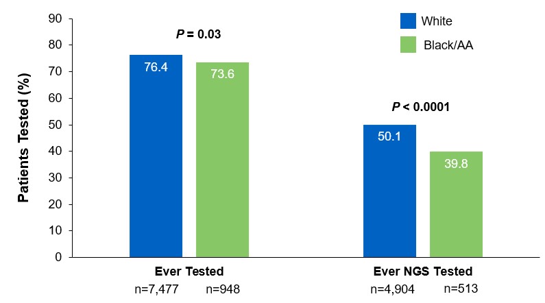
Beyond the underutilization of biomarker testing, there remains an even greater need to increase broad molecular testing among racial and ethnic minority groups in the US.12,13 In one retrospective study, Black/African American patients with advanced NSCLC had significantly lower rates of testing with NGS assays (39.8%) compared with White patients (50.1%) (Figure 3).12

††A retrospective study assessing real-world biomarker testing patterns in patients with de novo mNSCLC (N=2,257) in the community oncology setting using the US Oncology Network electronic health records between January 1st, 2017 and September 31st, 2019 with follow-up through December 31st, 2019.11

Figure 2: MYLUNG Consortium™ EMR Analysis of Patients With Metastatic NSCLC7,‡‡MYLUNG-Consortium‡‡A retrospective, observational study assessing real-world biomarker testing patterns in patients with metastatic NSCLC(N=3,474) from community oncology practices within the US Oncology Network community practices between 2018 and 2020.7
Figure 3: EMR Analysis of Biomarker Testing in Patients With Advanced/Metastatic NSCLC12,§§
EMR-Analysis-of-Biomarker-Testing
§§From a retrospective cohort study of patients with advanced/metastatic: NSCLC (N=14,768) from ~800 sites of care identified via the Flatiron Electronic Health Record Database between 2017 and 2020. Of this study cohort, patients included White (n=9,793), Black/African American (n=1,288), and non-squamous NSCLC (n=10,333).

Collectively, these findings highlight the disparity in proactive disease management across different patient populations.7,11,12

Considerations Across the Biomarker Testing Journey

There are several different methods in which eligible patients can be tested for actionable genetic alterations, each with unique considerations as indicated below (Figure 4).

Figure 4: Comparing Biomarker Testing Methods and Sample Types
Comparing-Biomarker-Testing-Methods***Data from a review of common molecular assays for biomarker testing that analyzed common detected variants, sensitivities, and turnaround time.6 †††cfDNA refers to all circulating DNA (largely non-malignant), while ctDNA refers to the tumor-related component of cfDNA.15 ‡‡‡Data from a prospective study that enrolled patients with previously untreated metastatic NSCLC undergoing SOC tissue genotyping and comprehensive cfDNA analysis, with turnaround time defined as the number of days between test order date and the retrieval of test results.16

While tissue biopsy remains the “gold standard” in NSCLC, it may not be feasible (insufficient tissue) or pragmatic (urgent need to begin treatment) in all patients.17 Plasma ctDNA demonstrates complementary results to tissue-based assays and can be considered a valid tool for genotyping of newly diagnosed patients with advanced NSCLC.15 In a prospective study of patients with previously untreated, non-squamous metastatic NSCLC from 2016 to 2018, guideline-recommended biomarkers with FDA-approved therapies (EGFR Exon 19 deletion and L858R, ALK fusion, ROS1 fusion, BRAF V600E) showed ≥ 98.2% concordance between tissue and liquid-based testing.16 While concordance is high for any single test, high concordance for full panels will be required for liquid biopsies to become standard; additionally, negative results on liquid biopsy still require validation with tissue testing.16,17

Liquid biopsy may offer improvements in sample acquisition and small tissue samples and provides less invasive procedures and shortened turnaround times.17 Other considerations for maximizing the tissue journey include the use of comprehensive testing, rapid on-site evaluation (ROSE), and implementing reflex testing protocols with the help of a multidisciplinary team (MDT).17

Delays in Biomarker Testing Results May Impact Treatment Plan Decisions18

Longer turnaround times for molecular testing compared with turnaround times for PD-L1 testing by IHC may result in the initiation of immunotherapy before molecular testing results are received.18 Waiting for complete biomarker test results prior to initiating therapy can allow doctors to make the most informed decisions surrounding a patient’s treatment journey.18

Consider Comprehensive Biomarker Testing as an Important Part of Your Treatment Plan8

As the NSCLC landscape continues to progress with the increasing number of actionable biomarkers, there is a growing need for proactive and comprehensive molecular testing.7,17 Although real-world data has shown significant underuse of biomarker testing, rates can be improved with diligent observation of expanding guidelines and recommendations by expert panels and associations.7,8 In the coming years, clinicians may consider evolving institutional protocols, including enabling reflex testing, and work as an MDT to ensure biomarker testing is performed on all eligible patients with advanced NSCLC.17

[Abbreviations]
AA, African American; ALK, anaplastic lymphoma kinase; BRAF, proto-oncogene B-Raf; cfDNA, cell-free DNA; ctDNA, circulating tumor DNA; EGFR, epidermal growth factor receptor; EMR, electronic medical record; ERBB2, erb-b2 receptor tyrosine kinase 2; HER2, human epidermal growth factor receptor 2; IHC, immunohistochemistry; KRAS, Kirsten rat sarcoma viral oncogene homolog; MET, mesenchymal-to-epithelial transition; mNSCLC, metastatic non-small cell lung cancer; NSCLC, non-small cell lung cancer; NCCN, National Comprehensive Cancer Network; NGS, next-generation sequencing; NTRK, neurotrophic tyrosine receptor kinase; PD-L1, programmed cell death ligand 1; RET, rearranged during transfection; ROS1, c-ros oncogene 1; SOC, standard-of-care.

[References]
1. Majeed U, et al. J Hematol Oncol. 2021;14:108.
2. Sung H, et al. CA Cancer J Clin. 2021;71:209-249.
3. Siegel RL, et al. CA Cancer J Clin. 2021;71:7-33.
4. Food and Drug Administration. www.fda.gov. Accessed October 6, 2021.
5. Referenced with permission from the NCCN Clinical Practice Guidelines in Oncology (NCCN Guidelines®) for Non-Small Cell Lung Cancer. V.3.2022. ©National Comprehensive Cancer Network, Inc. 2022. All rights reserved. Accessed March 16, 2022. To view the most recent and complete version of the guideline, go online to NCCN.org. NCCN makes no warranties of any kind whatsoever regarding their content, use or application and disclaims any responsibility for their application or use in any way.
6. Pennell NA, et al. Am Soc Clin Oncol Educ Book. 2019;39:531-542.
7. Robert NJ, et al. Presented at: The American Society of Clinical Oncology Annual Meeting; June 4–8, 2021; Virtual Meeting. Abstract 102.
8. John A, et al. Adv Ther. 2021;38:1552-1566.
9. Hanna N, et al. J Clin Oncol. 2017;35:3484-3515.
10. Lindeman NI, et al. Arch Pathol Lab Med. 2018;142:321-346.
11. Nadler ES, et al. Presented at: The American Society of Clinical Oncology Annual Meeting; June 4–8, 2021; Virtual Meeting. Abstract 9079.
12. Bruno DS, et al. Presented at: The American Society of Clinical Oncology Annual Meeting; June 4–8, 2021; Virtual Meeting. Abstract 9005.
13. Hann KEJ, et al. BMC Public Health. 2017;17:503.
14. Pennell NA, et al. JCO Precis Oncol. 2019;3:1-9.
15. Rolfo C, et al. J Thorac Oncol. 2021;16:1647-1662.
16. Leighl NB, et al. Clin Cancer Res. 2019;25:4691-4700.
17. Gregg JP, et al. Transl Lung Cancer Res. 2019;8:286-301.
18. Smeltzer MP, et al. J Thorac Oncol. 2020;15:1434-1448.

USA-510-80864 02/22

Mutations of STK11/KRAS Genes and Efficacy of Immunotherapy in NSCLC

SUMMARY: The American Cancer Society estimates that for 2022, about 236,740 new cases of lung cancer will be diagnosed and 135,360 patients will die of the disease. Lung cancer is the leading cause of cancer-related mortality in the United States. Non-Small Cell Lung Cancer (NSCLC) accounts for approximately 85% of all lung cancers and Adenocarcinoma now is the most frequent histologic subtype of lung cancer.

Immune checkpoints are cell surface inhibitory proteins/receptors that are expressed on activated T cells. They harness the immune system and prevent uncontrolled immune reactions by switching off the T cells of the immune system. Immune checkpoint proteins/receptors include CTLA-4 (Cytotoxic T-Lymphocyte Antigen 4, also known as CD152) and PD-1(Programmed cell Death 1). Checkpoint inhibitors unleash the T cells resulting in T cell proliferation, activation, and a therapeutic response.

TECENTRIQ® (Atezolizumab) is an anti-PDL1 monoclonal antibody, designed to directly bind to PD-L1 expressed on tumor cells and tumor-infiltrating immune cells, thereby blocking its interactions with PD-1 and B7.1 receptors and thus enabling the activation of T cells. AVASTIN® (Bevacizumab) is a biologic antiangiogenic antibody, directed against Vascular Endothelial Growth Factor (VEGF), and prevents the interaction of VEGF to its receptors (Flt-1 and KDR) on the surface of endothelial cells. The interaction of VEGF with its receptors has been shown to result in endothelial cell proliferation and new blood vessel formation. Combining TECENTRIQ® and AVASTIN® is supported by the following scientific rationale. AVASTIN® in addition to its established anti-angiogenic effects, may further enhance the ability of TECENTRIQ® to restore anti-cancer immunity, by inhibiting VEGF-related immunosuppression, promoting T-cell tumor infiltration and enabling priming and activation of T-cell responses against tumor antigens.

IMpower150 is a multicenter, open-label, randomized, Phase III study, conducted to evaluate the efficacy and safety of TECENTRIQ® in combination with Carboplatin and Paclitaxel with or without AVASTIN®, in patients with Stage IV, treatment naïve, non-squamous NSCLC. This study enrolled 1,202 patients, who were randomized (1:1:1) to receive either TECENTRIQ® along with Carboplatin and Paclitaxel (ACP-Group A), TECENTRIQ® and AVASTIN® along with Carboplatin and Paclitaxel (ABCP-Group B), or AVASTIN® plus Carboplatin and Paclitaxel (BCP-Group C – control arm). During the treatment-induction phase, patients in Group A received TECENTRIQ® 1200 mg IV along with Carboplatin AUC 6 and Paclitaxel 200mg/m2 IV on Day 1 of a 3-week treatment cycle for 4 or 6 cycles. Following the induction phase, patients received maintenance treatment with TECENTRIQ® on the same dose schedule until disease progression. Patients in Group B received AVASTIN® 15 mg/kg IV, along with TECENTRIQ®, Carboplatin and Paclitaxel IV, Day 1 of a 3-week treatment cycle for 4 or 6 cycles followed by maintenance treatment with the TECENTRIQ® and AVASTIN® until disease progression. Patients in the control Group C received AVASTIN® plus Carboplatin and Paclitaxel every 3 weeks for 4 or 6 cycles followed by AVASTIN® maintenance treatment until disease progression. Among randomized patients with tumors demonstrating no ALK and EGFR mutations, ABCP was associated with significant improvements in Progression Free Survival (PFS) and Overall Survival (OS), compared with BCP, in an updated OS analysis. ABCP also prolonged OS and PFS compared with BCP in an exploratory subgroup analysis of patients with EGFR-sensitizing mutations.

The Serine‐Threonine Kinase 11 (STK11) gene is located on the short arm of chromosome 19 and germline STK11 mutations are often detected in Peutz‐Jeghers syndrome, an Autosomal Dominant disorder resulting in mucocutaneous hyperpigmentation, hamartomas throughout the gastrointestinal tract, and a predisposition for breast, lung, pancreas, and gastrointestinal malignancies including cancers of the colon and small bowel. Both STK11 (also called LKB1) and KEAP1 mutation occur in about 17% of NSCLC (adenocarcinomas), respectively, and correlates with poor outcome with immune checkpoint inhibitors or immune checkpoint inhibitors plus chemotherapy. Although immune checkpoint inhibitors with or without chemotherapy have demonstrated survival benefit in patients with KRAS mutated tumors, it remains unclear how co-occurring STK11, KEAP1, and TP53 mutations affect outcomes following immune checkpoint blockade.

The authors in this publication conducted a retrospective exploratory analysis of the efficacy of ABCP (TECENTRIQ® and AVASTIN® along with Carboplatin and Paclitaxel), in patients with KRAS mutations and co-occuring STK11, KEAP1, or TP53 mutations, from the IMpower150 nonsquamous NSCLC patient population. Mutation status was determined by circulating tumor DNA Next-Generation Sequencing.

Among the KRAS mutated population, there was numerical improvement in median OS with ABCP compared to BCP (19.8 vs 9.9 months; HR=0.50), as well as PFS (8.1 vs 5.8 months; HR=0.42) respectively. The median OS with ACP (TECENTRIQ® along with Carboplatin and Paclitaxel) was 11.7 vs 9.9 months (HR=0.63), and PFS was 4.8 vs 5.8 months (HR=0.80), when compared with BCP (AVASTIN® plus Carboplatin and Paclitaxel). When compared to BCP, the ABCP group showed numerically greater survival than the ACP group among KRAS mutated patients. These results were consistent with reported survival improvements with immune checkpoint inhibitors in KRAS-mutant NSCLC.

In KRAS mutant patients across PD-L1 subgroups, OS and PFS were longer with ABCP when compared with BCP, but in PD-L1-low and PD-L1-negative subgroups, OS with ACP was similar to BCP. Conversely, in KRAS wild type patients, OS was longer with ACP than with ABCP or BCP across PD-L1 subgroups.

KRAS was frequently comutated with STK11, KEAP1, and TP53 and these subgroups conferred different prognostic outcomes. Within the KRAS mutated population, STK11 and/or KEAP1 mutations were associated with inferior OS and PFS across treatments compared with STK11-wild type and/or KEAP1wild type. In KRAS mutated patients with co-occurring STK11 and/or KEAP1 mutations (44.9%) or TP53 mutations (49.3%), survival was longer with ABCP than with ACP or BCP.

It was concluded that this analysis supported previous findings of mutation of STK11 and/or KEAP1 as poor prognostic indicators. Even though the clinical efficacy of ABCP (TECENTRIQ® and AVASTIN® along with Carboplatin and Paclitaxel) and ACP (TECENTRIQ® along with Carboplatin and Paclitaxel) was favorable compared with BCP (AVASTIN® plus Carboplatin and Paclitaxel) in these mutational subgroups, survival benefits were greater in the KRAS mutated and KEAP1 and STK11 wild type population versus KRAS mutated and KEAP1 and STK11 mutated population, suggesting both prognostic and predictive value of mutational analysis. The researchers added that these results suggest that TECENTRIQ® in combination with AVASTIN® and chemotherapy is an efficacious first-line treatment in metastatic NSCLC subgroups with KRAS mutations co-occurring with STK11 and/or KEAP1 or TP53 mutations and/or high PD-L1 expression.

Clinical efficacy of atezolizumab plus bevacizumab and chemotherapy in KRAS- mutated non-small cell lung cancer with STK11, KEAP1, or TP53 comutations: subgroup results from the phase III IMpower150 trial. West JH, McCleland M, Cappuzzo, F, et al. J Immunother Cancer. 2022 Feb;10(2):e003027. doi: 10.1136/jitc-2021-003027.

TIBSOVO® and VIDAZA® Combo Improve Survival in IDH1-Mutated Acute Myeloid Leukemia

SUMMARY: The American Cancer Society estimates that for 2022, about 20,050 new cases of Acute Myeloid Leukemia (AML) will be diagnosed in the United States and 11,540 patients will die of the disease. AML can be considered as a group of heterogeneous diseases with different clinical behavior and outcomes. Cytogenetic analysis has been part of routine evaluation when caring for patients with AML. By predicting resistance to therapy, tumor cytogenetics will stratify patients, based on risk and help manage them accordingly. Even though cytotoxic chemotherapy may lead to long term remission and cure in a minority of patients with favorable cytogenetics, patients with high risk features such as unfavorable cytogenetics, molecular abnormalities, prior Myelodysplasia and advanced age, have poor outcomes with conventional chemotherapy alone. AML mainly affects older adults and the median age at diagnosis is 68 years. A significant majority of patients with AML are unable to receive intensive induction chemotherapy due to comorbidities and therefore receive less intensive, noncurative regimens, with poor outcomes.

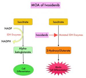
Isocitrate DeHydrogenase (IDH) is a metabolic enzyme that helps generate energy from glucose and other metabolites, by catalyzing the conversion of Isocitrate to Alpha-Ketoglutarate. Alpha-ketoglutarate is required to properly regulate DNA and histone methylation, which in turn is important for gene expression and cellular differentiation. IDH mutations lead to aberrant DNA methylation and altered gene expression thereby preventing cellular differentiation, with resulting immature undifferentiated cells. IDH mutations can thus promote leukemogenesis in Acute Myeloid Leukemia and tumorigenesis in solid tumors and can result in inferior outcomes. There are three isoforms of IDH. IDH1 is mainly found in the cytoplasm, as well as in peroxisomes, whereas IDH2 and IDH3 are found in the mitochondria, and are a part of the Krebs cycle. Approximately 20% of patients with AML, 70% of patients with Low-grade Glioma and secondary Glioblastoma, 50% of patients with Chondrosarcoma, 20% of patients with Intrahepatic cholangiocarcinoma, 30% of patients with Angioimmunoblastic T-cell lymphoma and 8% of patients with Myelodysplastic syndromes/Myeloproliferative neoplasms, are associated with IDH mutations.MOA-of-Ivosidenib

TIBSOVO® (Ivosidenib) is a first-in-class, oral, potent, targeted, small-molecule inhibitor of mutant IDH1. The FDA in 2018, approved TIBSOVO® for adult patients with relapsed or refractory AML with a susceptible IDH1 mutation and in 2019 approved TIBSOVO® for newly diagnosed AML with a susceptible IDH1 (Isocitrate DeHydrogenase-1) mutation, in patients who are at least 75 years old or who have comorbidities that preclude the use of intensive induction chemotherapy. VIDAZA® (Azacitidine) is a hypomethylating agent that promotes DNA hypomethylation by inhibiting DNA methyltransferases. VIDAZA® has been shown to significantly improve Overall Survival (OS) when compared to conventional care regimens in elderly unfit patients with newly diagnosed AML, who are not candidates for intensive chemotherapy. In a Phase Ib trial, TIBSOVO® in combination with VIDAZA® showed encouraging clinical activity in newly diagnosed IDH1-mutated AML patients.

AGILE is a global, double-blind, randomized, placebo-controlled, Phase III trial in which the efficacy and safety of a combination of TIBSOVO® and VIDAZA® were assessed, as compared with placebo and VIDAZA®, in patients with newly diagnosed IDH1-mutated Acute Myeloid Leukemia, who were ineligible for intensive induction chemotherapy. Patients were randomly assigned in a 1:1 ratio to receive TIBSOVO® 500 mg orally once daily combined with VIDAZA® 75 mg/m2 subcutaneously or IV for 7 days in 28-day cycles (N=72) or placebo and VIDAZA® (N=74). All the patients were to be treated for a minimum of six cycles until disease progression or unacceptable toxicities. The median patient age was 76 years, 75% had primary AML and 25% had secondary AML, 67% had intermediate cytogenetic risk and 22% had poor cytogenetic risk. Patients were stratified according to geographic region and disease status (Primary versus Secondary Acute Myeloid Leukemia). The Primary end point was Event-Free Survival, defined as the time from randomization until treatment failure (failure of complete remission by week 24), relapse from remission, or death from any cause, whichever occurred first.

At a median follow-up of 12.4 months, Event-Free Survival was significantly longer in the TIBSOVO® and VIDAZA® group than in the placebo and VIDAZA® group (HR=0.33; P=0.002). This benefit was seen across all key subgroups. The estimated probability that a patient would remain event-free at 12 months was 37% in the TIBSOVO® and VIDAZA® group and 12% in the placebo and VIDAZA® group. The median Overall Survival was 24.0 months with TIBSOVO® and VIDAZA® and 7.9 months with placebo and VIDAZA® (HR=0.44; P=0.001). Among those patients who were dependent on transfusion of red blood cells, platelets, or both at baseline, a higher percentage of patients converted to transfusion independence with TIBSOVO® and VIDAZA®, than with placebo and VIDAZA® (46% versus 18%; P=0.006). Health-Related Quality of Life scores favored TIBSOVO® and VIDAZA® across all subscales. Grade 3 or higher Adverse Events included febrile neutropenia (28% with TIBSOVO® and VIDAZA® versus 34% with placebo and VIDAZA®) and neutropenia (27% versus 16%, respectively). Differentiation syndrome of any grade occurred in 14% of the patients receiving TIBSOVO® and VIDAZA® versus 8% among those receiving placebo and VIDAZA®.

It was concluded that a combination of TIBSOVO® and VIDAZA® significantly improved Event-Free Survival, Response Rates, and Overall Survival, as compared with placebo and VIDAZA®, in patients with newly diagnosed IDH1-mutated Acute Myeloid Leukemia, who were ineligible for induction chemotherapy. The authors added that treatment with TIBSOVO® and VIDAZA® resulted in better Quality of Life and higher rates of transfusion independence.

Ivosidenib and Azacitidine in IDH1-Mutated Acute Myeloid Leukemia. Montesinos P, Recher C, Vives S, et al. N Engl J Med 2022; 386:1519-1531.

Overall Survival at 2 Years with LUMAKRAS® for KRAS G12C Positive Non Small Cell Lung Cancer

SUMMARY: The American Cancer Society estimates that for 2022, about 236,740 new cases of lung cancer will be diagnosed and 135,360 patients will die of the disease. Lung cancer is the leading cause of cancer-related mortality in the United States. Non-Small Cell Lung Cancer (NSCLC) accounts for approximately 85% of all lung cancers. Of the three main subtypes of NSCLC, 30% are Squamous Cell Carcinomas (SCC), 40% are Adenocarcinomas and 10% are Large Cell Carcinomas. With changes in the cigarette composition and decline in tobacco consumption over the past several decades, Adenocarcinoma now is the most frequent histologic subtype of lung cancer.

The KRAS (kirsten rat sarcoma viral oncogene homologue) proto-oncogene encodes a protein that is a member of the small GTPase super family. The KRAS gene provides instructions for making the KRAS protein, which is a part of a signaling pathway known as the RAS/MAPK pathway. By relaying signals from outside the cell to the cell nucleus, the protein instructs the cell to grow, divide and differentiate. The KRAS protein is a GTPase, and converts GTP into GDP. To transmit signals, the KRAS protein must be turned on, by binding to a molecule of GTP. When GTP is converted to GDP, the KRAS protein is turned off or inactivated, and when the KRAS protein is bound to GDP, it does not relay signals to the cell nucleus. The KRAS gene is in the Ras family of oncogenes, which also includes two other genes, HRAS and NRAS. When mutated, oncogenes have the potential to change normal cells cancerous.

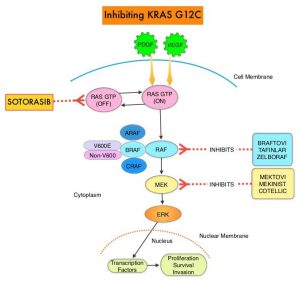
KRAS is the most frequently mutated oncogene in human cancers and are often associated with resistance to targeted therapies and poor outcomes. The KRAS-G12C mutation occurs in approximately 12-15% of Non Small Cell Lung Cancers (NSCLC) and in 3-5% of colorectal cancers and other solid cancers. KRAS G12C is one of the most prevalent driver mutations in NSCLC and accounts for a greater number of patients than those with ALK, ROS1, RET, and TRK 1/2/3 mutations combined. KRAS G12C cancers are genomically more heterogeneous and occur more frequently in current or former smokers, and are likely to be more complex genomically than EGFR mutant or ALK rearranged cancers. G12C is a single point mutation with a Glycine-to-Cysteine substitution at codon 12. This substitution favors the activated state of KRAS, resulting in a predominantly GTP-bound KRAS oncoprotein, amplifying signaling pathways that lead to oncogenesis.Inhibiting-KRAS-G12C

LUMAKRAS® (Sotorasib) is a first-in-class small molecule that specifically and irreversibly inhibits KRAS-G12C and traps KRAS-G12C in the inactive GDP-bound state. Preclinical studies in animal models showed that LUMAKRAS® inhibited nearly all detectable phosphorylation of Extracellular signal-Regulated Kinase (ERK), a key downstream effector of KRAS, leading to durable complete regression of KRAS-G12C tumors. The CodeBreaK clinical development program for LUMAKRAS® was designed to treat patients with an advanced solid tumor with the KRAS G12C mutation and address the longstanding unmet medical need for these cancers. This program has enrolled more than 800 patients across 13 tumor types since its inception.

CodeBreaK 100 is a Phase I and II, first-in-human, open-label, single arm, multicenter study, which enrolled patients with KRAS G12C-mutant solid tumors. Eligible patients must have received a prior line of systemic anticancer therapy, for their tumor type and stage of disease. The Phase II trial enrolled 126 patients with NSCLC, who had locally advanced or metastatic NSCLC with a KRAS G12C mutation, and had progressed on an immune checkpoint inhibitor and/or platinum-based chemotherapy. Patients with active brain metastases were excluded. Patient received LUMAKRAS® 960 mg orally once daily, until disease progression or unacceptable toxicity. The median age was 64 years, 52% were male, over 90% of patients had a smoking history, median number of prior lines of therapy was two, 92% had prior platinum-based chemotherapy and 90% had prior anti–PD-L1 therapy, 83% had both prior platinum-based chemotherapy and immunotherapy. The Primary end point of the trial was Overall Response Rate (ORR) as assessed by blinded Independent Central Review. Secondary end points included Duration of Response (DOR), Disease Control Rate (DCR), time to recovery, Progression Free Survival (PFS), Overall Survival (OS), and Safety. The examination of biomarkers served as an exploratory end point.

At the time of Primary analysis, at a median follow up of 12.2 months, the ORR was 37.1% and the median Duration of Response was 10 months. Based on the data from the primary analysis, the FDA in 2021 granted accelerated approval to LUMAKRAS®, for the treatment of patients with locally advanced or metastatic NSCLC, whose tumors harbor the KRAS G12C mutation, and who have received prior therapies.

For this updated analysis, the median follow up time for OS was 24.9 months, and the researchers included 174 patients enrolled in Phase I (N=48) and Phase II (N=126) portions of the CodeBreaK 100 trial, who were treated with LUMAKRAS®. The Overall Response Rate was 40.7% and the Disease Control Rate (DCR) was 83.7%. The median time to response was 6 weeks, the median Duration of Response was 12.3 month and 50.6% of responders remained in response for 12 months or more. The median PFS was 6.3 months and the median OS showed no change in the updated analysis, and was 12.5 months. At 1-year, the OS rate was 50.8% and the 2-year Overall Survival was 32.5%. The researchers performed additional analyses on both tumor and blood samples to identify biomarker profiles associated with durable clinical benefit and these showed that prolonged clinical benefit was observed regardless of Tumor Mutation Burden, PDL1 expression, and STK11 co-mutation status. Grade 3 or 4 treatment-related Adverse Events occurred in 21% of patients. Most adverse events were Grade 1 or 2, and treatment-related adverse events occurring in more than 10% of patients included diarrhea, elevated liver enzymes, nausea and fatigue.

It was concluded from this updated analysis that this is the longest follow up of patients on any KRAS G12C inhibitor, and LUMAKRAS® demonstrated meaningful and durable efficacy in patients with KRAS mutated NSCLC for whom treatment options are limited, following progression on first line treatment, and historically have had poor outcomes. Patients on LUMAKRAS® benefitted regardless of Tumor Mutation Burden, PDL1 expression, and STK11 co-mutation status. A global Phase III study (CodeBreaK 200) is underway, comparing LUMAKRAS® to Docetaxel in patients with KRAS G12C-mutated NSCLC.

Long-term outcomes with sotorasib in pretreated KRASp.G12C-mutated NSCLC: 2-year analysis of CodeBreaK100. Dy GK, Govindan R, Velcheti V, et al. Presented at: 2022 AACR Annual Meeting; April 8-13, 2022, New Orleans, LA. Abstract CT008.

Early-Stage Multi-Cancer Detection Using an Extracellular Vesicle Protein-Based Blood Test

SUMMARY: The American Cancer Society’s estimates that in 2022, about 62,210 people will be diagnosed with pancreatic cancer and 49,830 people will die of the disease, 19,880 women will receive a new diagnosis of ovarian cancer and about 12,810 women will die of the disease, and about 81,800 new cases of bladder cancer will be diagnosed in 2022 and about 17,100 patients will die of the disease. These three cancer types are estimated to account for approximately 80,000 deaths in the US in 2022. Detecting cancer at early stages can significantly increase survival rates and outcomes.

Several multi-cancer early detection tests are being developed that involve blood-based circulating cell-free tumor DNA (cfDNA) in the plasma, to track hundreds of patient-specific mutations, to detect Minimal Residual Disease (MRD) , as well as detection of abnormal methylation patterns, followed by machine learning approaches, to differentiate between cancer and non-cancer, for detecting clinically significant, late-stage (III and IV) cancers. Early detection of cancer however is the key to improving survival. This is particularly relevant for certain cancer types. Pancreatic Ductal AdenoCarcinoma (PDAC) is one of the deadliest cancers, and a leading cause of all cancer-related deaths in the United States, and is typically detected when the disease is advanced. However, when detected at Stage I, survival rates can be as high as 80%. Ovarian cancer is often detected when the disease is advanced and the 5-year survival rates are less than 30%, but can be as high as 93% when detected early. The same holds true for metastatic bladder cancer, with 5-year survival rates of only 6%, whereas while detected when the tumor is still localized to the bladder wall inner layer results in a 5-year survival rate of 96%. Even though serum CA19-9 is intended as an aid in the management of patients with confirmed pancreatic cancer for serial monitoring of their response to therapy and disease progression, it is not recommended by the FDA for screening, as it may be elevated in several benign conditions. Similarly, serum CA-125 is FDA approved for use in monitoring patients with ovarian cancer for disease persistence and recurrence, but is not recommended to screen for ovarian cancer. Currently, there are few general screening strategies to detect asymptomatic, early-stage PDAC, ovarian, or bladder cancer and there is therefore a significant unmet need in this patient group.

Exosomes are 30-150 nm-sized Extracellular Vesicles (EVs) secreted by multiple different cell types and ejected by tumors into the bloodstream. They mediate intercellular signaling by transferring mRNAs and microRNAs between distant cells and tissues and therefore carry functional protein biomarkers representing the tumor proteome. Exosomes represent one potential approach for more sensitive detection of cancer-related biomarkers from blood.

The researchers in this study used an Alternating Current Electrokinetic (ACE)-based platform (Verita™ System) to efficiently isolate EVs from soluble contaminants such as cells, small proteins, or other vesicles from patient samples, and then measured the concentrations of associated protein biomarkers (“EV proteins”) present in the purified EV samples from our case-control study subjects. The researchers chose this platform over the current gold standard ultracentrifugation method, which the authors felt was inefficient and not suitable for point-of-care applications. Artificial Intelligence machine-learning algorithm developed by the researchers, enabled detection of early-stage pancreatic, ovarian, and bladder cancers.

In this case-control pilot study, 139 pathologically confirmed Stage I and II cancer cases representing pancreatic, ovarian, or bladder patients were compared with 184 control subjects, using the Verita™ System. The Extracellular Vesicles (EVs) isolated using this technology, were consistent with the presence of Exosomes, in accordance with the International Society for Extracellular Vesicles (ISEV) 2018 guidelines. The researchers selected a panel of 13 Extracellular Vesicle (EV) proteins along with age, a known cofactor in cancer. In order to simulate a real-world screening scenario, all cancer cases were treatment-naïve and to ensure that these were early-stage patients, the histopathologic staging was confirmed using the American Joint Commission on Cancer (AJCC) guidelines. The median age of the cancer cases was 60 years and 63.3% of the overall cancer cases were Stage I, with the remaining 36.7% at Stage II. The median age of the control group was 57 years and had no known history of cancer, autoimmune diseases, neurodegenerative disorders or diabetes mellitus.

When the overall cancer case cohort was compared with the control individuals using the EV protein biomarker test, the average sensitivity was 71.2%, at a specificity of 99.5%. When considered across all the three cancers studied, EV protein biomarker test using this technology demonstrated similar sensitivities of 70.5% and 72.5% for Stage I and II patients, respectively. This new technology detected 95.5% of Stage I pancreatic cancers, 73.1% of pathologic Stage IA lethally aggressive serous ovarian adenocarcinomas and 43.8% in bladder cancer, demonstrating the potential value of this platform for detection of early stage cancers. The lower sensitivity for detecting early stage bladder cancer may be due to high molecular and histologic heterogeneity of bladder tumors.

It was concluded from this study that blood-based EV protein detection test has potential clinical value for early cancer detection and the use of Verita™ platform resulted in the accurate detection of early stage pancreatic, ovarian, or bladder cancer. The authors added that mortality from pancreatic cancer which will soon become the second leading cause of cancer mortality in the U.S., can be greatly reduced if this study results are validated.

Early-stage multi-cancer detection using an extracellular vesicle protein-based blood test. Hinestrosa, J.P., Kurzrock, R., Lewis, J.M. et al. Commun Med 2, 29 (2022). https://doi.org/10.1038/s43856-022-00088-6.

FDA Approves PSMA Targeted Therapy for Metastatic Castrate Resistant Prostate Cancer

SUMMARY: The FDA on March 23, 2022, approved PLUVICTO® (Lutetium Lu 177 vipivotide tetraxetan) for the treatment of adult patients with Prostate-Specific Membrane Antigen (PSMA)-positive metastatic Castration-Resistant Prostate Cancer (mCRPC), who had been treated with Androgen Receptor (AR) pathway inhibition and Taxane-based chemotherapy. The FDA also approved LOCAMETZ® (Gallium Ga 68 gozetotide), a radioactive diagnostic agent for Positron Emission Tomography (PET) of PSMA-positive lesions, including selection of patients with metastatic prostate cancer for whom PLUVICTO® PSMA-directed therapy is indicated. LOCAMETZ® is the first radioactive diagnostic agent approved for patient selection in the use of a radioligand therapeutic agent.

The development and progression of prostate cancer is driven by androgens. Androgen Deprivation Therapy (ADT) or testosterone suppression has therefore been the cornerstone of treatment of advanced prostate cancer, and is the first treatment intervention. Approximately 10-20% of patients with advanced Prostate cancer will progress to Castration Resistant Prostate Cancer (CRPC) within five years during ADT, and over 80% of these patients will have metastatic disease at the time of CRPC diagnosis. Among those patients without metastases at CRPC diagnosis, 33% are likely to develop metastases within two years. Progression to Castration Resistant Prostate Cancer (CRPC) often manifests itself with a rising PSA (Prostate Specific Antigen) and the estimated mean survival of patients with CRPC is 9-36 months, and there is therefore an unmet need for new effective therapies.

Prostate-Specific Membrane Antigen (PSMA) is a Type II cell membrane glycoprotein that is selectively expressed in prostate cells, with high levels of expression in prostatic adenocarcinoma. PSMA is a therefore an excellent target for molecular imaging and therapeutics, due to its high specificity for prostate cancer.

PLUVICTO® is a radiopharmaceutical that targets PSMA. It is comprised of Lutetium-177, a cytotoxic radionuclide , linked to the ligand PSMA-617, a small molecule designed to bind with high affinity to PSMA. Radioligand therapy with PLUVICTO® targets PSMA and releases its payload of lethal beta radiation into the prostate cancer cell. The antitumor activity and safety PLUVICTO® have been established previously in a Phase II study (Lancet Oncol. 2018;19:825-833).

VISION is an international, randomized, open-label Phase III study in which the benefit of PLUVICTO® was evaluated in men with PSMA-positive mCRPC, previously treated with second generation Androgen Receptor signaling pathway inhibitor (XTANDI® -Enzalutamide or ZYTIGA®-Abiraterone acetate), and 1 or 2 taxane chemotherapy regimens. In this trial, 831 patients were randomized 2:1 to receive PLUVICTO® 7.4 GBq (200 mCi) every 6 weeks for up to a total of 6 cycles plus Standard of Care as determined by the treating physician (N=551), or Standard of Care only (N=280). Both treatment groups were well balanced and this trial excluded patients treated with XOFIGO® (Radium-223). All enrolled patients received a GnRH analog or had prior bilateral orchiectomy and had a castrate level or serum/plasma testosterone of lower than 50ng/dL. PET imaging with LOCAMETZ® was used to determine PSMA positivity by central review. PSMA-positive mCRPC was defined as having at least one tumor lesion with LOCAMETZ® uptake greater than normal liver. Patients were excluded from enrollment if any lesions exceeding certain size criteria in the short axis had uptake less than or equal to uptake in normal liver. The Primary endpoints were radiographic Progression Free Survival (rPFS) by Independent Central Review (ICR) and Overall Survival (OS). Secondary endpoints included Objective Response Rate (ORR), Disease Control Rate (DCR), and time to first Symptomatic Skeletal Event (SSE). The median study follow up was 20.9 months.

Treatment with the combination of PLUVICTO® plus Standard of Care resulted in a statistically significant improvement in the Primary endpoints of Overall Survival and radiographic Progression Free Survival. PLUVICTO® plus Standard of Care significantly improved rPFS by 60%, compared to Standard of Care alone (median rPFS 8.7 versus 3.4 months, HR=0.40; P<0.001). The median OS was also significantly improved by 38% in the study group (median OS 15.3 versus 11.3 months, HR=0.62; P<0.001). All key secondary endpoints including Objective Response Rate, Disease Control Rate, and time to first Symptomatic Skeletal Event were statistically significant, and in favor of PLUVICTO® plus Standard of Care. The most common adverse reactions in patients receiving PLUVICTO® were fatigue, dry mouth, nausea, decreased appetite, constipation, anemia, lymphopenia, thrombocytopenia, hypocalcemia and hyponatremia.

It was concluded that radioligand therapy with PLUVICTO® significantly improved radiographic Progression Free Survival and Overall Survival when added to Standard of Care, compared with Standard of Care alone, in men with PSMA-positive metastatic Castration Resistant Prostate Cancer.

Lutetium-177-PSMA-617 for Metastatic Castration-Resistant Prostate Cancer. Sartor O, de Bono J, Chi KN, et al. N Engl J Med 2021; 385:1091-1103.

LYNPARZA® Superior to Next-Generation Hormonal Drug in CRPC Patients with Homologous Recombination Repair Gene Alterations

SUMMARY: Prostate cancer is the most common cancer in American men with the exclusion of skin cancer, and 1 in 9 men will be diagnosed with Prostate cancer during their lifetime. It is estimated that in the United States, about 268,490 new cases of Prostate cancer will be diagnosed in 2022 and 34,500 men will die of the disease. The development and progression of Prostate cancer is driven by androgens. Androgen Deprivation Therapy (ADT) or testosterone suppression has therefore been the cornerstone of treatment of advanced Prostate cancer and is the first treatment intervention. Approximately 10-20% of patients with advanced Prostate cancer will progress to Castration Resistant Prostate Cancer (CRPC) within five years during ADT, and over 80% of these patients will have metastatic disease at the time of CRPC diagnosis. The malignant transformation of prostatic epithelial cell as well as the development of CRPC has been attributed to deleterious alterations in a variety of genes including loss-of-function alterations in Homologous Recombination Repair (HRR) genes.

DNA damage is a common occurrence in daily life by UV light, ionizing radiation, replication errors, chemical agents, etc. This can result in single and double strand breaks in the DNA structure which must be repaired for cell survival. The two vital pathways for DNA repair in a normal cell are BRCA1/BRCA2 and PARP. BRCA1 and BRCA2 are tumor suppressor genes that recognize and repair double strand DNA breaks via Homologous Recombination Repair (HRR) pathway. Homologous Recombination is a type of genetic recombination, and is a DNA repair pathway utilized by cells to accurately repair DNA double-stranded breaks during the S and G2 phases of the cell cycle, and thereby maintain genomic integrity. Homologous Recombination Deficiency (HRD) is noted following mutation of genes involved in HR repair pathway. At least 15 genes are involved in the Homologous Recombination Repair (HRR) pathway including BRCA1, BRCA2, PALB2, CHEK2 and ATM genes. Mutations in these genes predispose an individual to develop malignant tumors. Mutations in BRCA1 and BRCA2 account for about 20-25% of hereditary breast cancers and about 5-10% of all breast cancers. They also account for 15% of ovarian cancers, in addition to other cancers such as Colon and Prostate. BRCA mutations can either be inherited (Germline) and present in all individual cells or can be acquired and occur exclusively in the tumor cells (Somatic). Somatic mutations account for a significant portion of overall BRCA1 and BRCA2 aberrations. Loss of BRCA function due to frequent somatic aberrations likely deregulates HR pathway, and other pathways then come in to play, which are less precise and error prone, resulting in the accumulation of additional mutations and chromosomal instability in the cell, with subsequent malignant transformation. Homologous Recombination Deficiency therefore indicates an important loss of DNA repair function.

The PARP (Poly ADP Ribose Polymerase), family of enzymes include, PARP1 and PARP2, and is a related enzymatic pathway that repairs single strand breaks in DNA. In a BRCA mutant, the cancer cell relies solely on PARP pathway for DNA repair to survive. PARP inhibitors trap PARP onto DNA at sites of single-strand breaks, preventing their repair and generating double-strand breaks that cannot be repaired accurately in tumors harboring defects in Homologous Recombination Repair pathway genes, such as BRCA1 or BRCA2 mutations, and this leads to cumulative DNA damage and tumor cell death.

LYNPARZA® (Olaparib) is a first-in-class PARP inhibitor and blocks DNA damage response in tumors harboring a deficiency in Homologous Recombination Repair, as is noted in those with mutations such as BRCA1 and/or BRCA2. LYNPARZA® showed promising results in a Phase II trial (TOPARP), when given as monotherapy, in patients with BRCA1/2 or ATM gene-mutated mCRPC, who had received a prior Taxane-based chemotherapy, and at least one newer hormonal agent (ZYTIGA® or XTANDI®).

PROfound is a prospective, multicentre, randomized, open-label, Phase III trial in which the efficacy and safety of LYNPARZA® was compared with physician’s choice of either XTANDI® or ZYTIGA® in two groups of patients with mCRPC, who had progressed on prior treatment with new hormonal anticancer treatments, and had a qualifying tumor mutation in one of 15 genes involved in the Homologous Recombination Repair (HRR) pathway. Patients in Cohort A (N=245) had alterations in BRCA1, BRCA2 or ATM genes while those in Cohort B (N=142) had alterations in any one of 12 other genes known to be involved in DNA repair which included BRIP1, BARD1, CDK12, CHEK1, CHEK2, FANCL, PALB2, PPP2R2A, RAD51B, RAD51C, RAD51D or RAD54L. Patients were randomized 2:1 within each cohort to receive LYNPARZA® 300 mg orally BID or physician’s choice of XTANDI® 160 mg orally QD or ZYTIGA® 1000 mg orally QD along with Prednisone 5 mg orally BID. Patient characteristics were well-balanced between arms in each treatment group, median patient age was 68 years, approximately 25% of patients had de novo metastatic disease, about 65% of patients received prior Taxane therapy and more than 20% had received two lines of chemotherapy. Patients were allowed to cross over to LYNPARZA® upon progression. The Primary endpoint was radiographic Progression-Free Survival (rPFS) in Cohort A, assessed by Blinded Independent Central Review (BICR).

The authors had previously reported that in Cohort A, the median PFS was 7.4 months with LYNPARZA®, compared to 3.5 months in the control group (HR=0.34, P<0.0001). This represented a 66% greater delay in disease progression compared to hormonal therapy. The interim Overall Survival analysis in Cohort A showed that median OS was 18.5 months with LYNPARZA® compared to 15 months with control drug treatment (HR=0.64, P=0.0173). In Cohort A, the Objective Response Rate (ORR) was 33.3% with LYNPARZA® compared with 2.3% with control drug therapies (P<0.0001).

The authors in this publication reported the results of the prespecified Secondary endpoints, which included pain, Health-Related Quality of Life (HRQOL), symptomatic Skeletal-Related Events, and time to first opiate use for cancer-related pain in Cohort A group of patients. Pain was assessed with the Brief Pain Inventory-Short Form, and HRQOL was assessed with the Functional Assessment of Cancer Therapy-Prostate (FACT-P). Cohort A included 245 patients with alterations in BRCA1, BRCA2, or ATM genes, of whom 162 patients received the investigational agent LYNPARZA®, and 83 patients received control drug. The median duration of follow up at data cutoff was 6.2 months for all LYNPARZA® group patients and 3.5 months for the control group patients.

The median time to pain progression was significantly longer with LYNPARZA® and was Not Reached in the LYNPARZA® group versus 9.92 months in the control group (HR=0.44; P=0.019). Pain interference scores were also significantly better in the LYNPARZA® group (difference in overall adjusted mean change from baseline score −0.85; nominal P=0.0004). Median time to progression of pain severity was Not Reached in either group. Among patients who had not used opiates at baseline (113 in the LYNPARZA® group, 58 in the control group), median time to first opiate use for cancer-related pain was 18.0 months in the LYNPARZA® group versus 7.5 months in the control group (HR=0.61; nominal P=0.044).

The proportion of patients with clinically meaningful improvement in FACT-P total score during treatment was higher for the LYNPARZA® group than the control group (10% versus 1% respectively; odds ratio=8.32; nominal P=0.0065). The median time to first symptomatic Skeletal-Related Event was not reached for either treatment group and the proportions of patients remaining free of symptomatic Skeletal-Related Events were 89.5% versus 77.1% at 6 months and 77.6% versus 53.6% at 12 months, in the LYNPARZA® and control groups respectively.

It was concluded that LYNPARZA® was associated with reduced pain burden and better-preserved HRQOL compared with the two control drugs, in patients with metastatic Castration-Resistant Prostate Cancer and Homologous Recombination Repair gene alterations, who had disease progression after a previous next-generation hormonal drug. The authors added that the study findings support the clinical benefit of improved radiographical Progression Free Survival and Overall Survival identified in PROfound trial.

Pain and health-related quality of life with olaparib versus physician’s choice of next-generation hormonal drug in patients with metastatic castration-resistant prostate cancer with homologous recombination repair gene alterations (PROfound): an open-label, randomised, phase 3 trial. Thiery-Vuillemin A, de Bono J, Hussain M, et al. Published:February 11, 2022. DOI:https://doi.org/10.1016/S1470-2045(22)00017-1

Cancer Risks Associated With BRCA1 and BRCA2 Pathogenic Variants

SUMMARY: DNA damage is a common occurrence in daily life by UV light, ionizing radiation, replication errors, chemical agents, etc. This can result in single and double strand breaks in the DNA structure which must be repaired for cell survival. The vital pathways for DNA repair in a normal cell are BRCA1/BRCA2 and PARP. BRCA1 and BRCA2 genes recognize and repair double strand DNA breaks via Homologous Recombination Repair (HRR) pathway. Homologous Recombination is a type of genetic recombination, and is a DNA repair pathway utilized by cells to accurately repair DNA double-stranded breaks during the S and G2 phases of the cell cycle, and thereby maintain genomic integrity. Homologous Recombination Deficiency (HRD) is noted following mutation of genes involved in HR repair pathway.

BRCA1 and BRCA2 are tumor suppressor genes located on chromosome 17 and chromosome 13 respectively and functional BRCA proteins repair damaged DNA, and play an important role in maintaining cellular genetic integrity. They regulate cell growth and prevent abnormal cell division and development of malignancy. Mutations in these genes predispose an individual to develop malignant tumors. It is well established that the presence of BRCA1 and BRCA2 mutations can significantly increase the lifetime risk for developing breast and ovarian cancer, as high as 85% and 40% respectively.

BRCA mutations can either be inherited (Germline) and present in all individual cells or can be acquired and occur exclusively in the tumor cells (Somatic). Somatic mutations account for a significant portion of overall BRCA1 and BRCA2 aberrations. Loss of BRCA function due to frequent somatic aberrations likely deregulates HR pathway, and other pathways then come in to play, which are less precise and error prone, resulting in the accumulation of additional mutations and chromosomal instability in the cell, with subsequent malignant transformation. Homologous Recombination Deficiency therefore indicates an important loss of DNA repair function.

Pathogenic Variants (PVs) in BRCA1 and BRCA2 (BRCA1/2) are well known to be associated with increased lifetime risk for breast and ovarian cancer in women, and reliable risk estimates are also available and can be as high as 85% and 40% respectively. However, the association of BRCA1 and BRCA2 Pathogenic Variants with cancers other than female breast and ovarian cancers remain uncertain, and these associations have been based on studies with relatively small sample sizes, resulting in imprecise cancer risk estimates. It is therefore important that precise risk estimates are available, in order to optimize clinical management strategies and guidelines for cancer risk management in female and male BRCA1/2 carriers. The NCCN and other guidelines recommend breast and ovarian cancer screening for BRCA1/2 carriers, prostate cancer screening for BRCA2 carriers. Screening is also recommended for pancreatic cancer in BRCA1/2 carriers, but only in the presence of a positive family history of the disease.

The authors conducted this analysis to provide comprehensive and precise age-specific risk estimates of 22 cancers other than female breast and ovarian cancers associated with Pathogenic Variants in BRCA1 and BRCA2, for effective cancer risk management. The researchers used data from 3,184 BRCA1 families and 2,157 BRCA2 families in the Consortium of Investigators of Modifiers of BRCA1/2 (CIMBA), to estimate age-specific Relative Risk (RR) and absolute risks for 22 first primary cancer types, after adjusting for family ascertainment. CIMBA was formed by a collaborative group of researchers working on genetic modifiers of cancer risk in BRCA1 and BRCA2 mutation carriers and provides sufficient sample sizes to allow large scale studies, in order to reliably evaluate the effects of genetic modifiers.

BRCA1 Pathogenic Variants were associated with significantly increased risk of male breast cancer (RR = 4.30; 4.3 times increased risk), pancreatic cancer (RR = 2.36), and stomach cancer (RR = 2.17). Although associations of BRCA1 Pathogenic Variants with colorectal and gallbladder cancers were observed, the results were not robust in the sensitivity analyses performed.

BRCA2 Pathogenic Variants were associated with increased risk of male breast cancer (RR = 44.0), stomach cancer (RR = 3.69), pancreatic cancer (RR = 3.34) and prostate cancer (RR = 2.22). Female carriers had a higher risk of stomach cancer than male carriers (RR = 6.89 versus 2.76; P=0.04).

The absolute/cumulative risks to age 80 years ranged from 0.4% for male breast cancer to approximately 2.5% for pancreatic cancer for BRCA1 carriers and from approximately 2.5% for pancreatic cancer to 27% for prostate cancer for BRCA2 carriers. In the present study, previously suggested associations of BRCA1 Pathogenic Variants with risks of other genitourinary cancers and increased risk of bone, brain, blood, gallbladder cancers and melanoma for BRCA2 Pathogenic Variants, were not replicated.

It was concluded from this analysis that in addition to female breast and ovarian cancers, BRCA1 and BRCA2 Pathogenic Variants are associated with increased risks of male breast cancer, pancreatic cancer, stomach cancer, and prostate cancer, the later only with BRCA2 Pathogenic Variants , but are not associated with the risks of other previously suggested cancers. These findings provide age-specific cancer risk estimates and will allow for improved cancer risk assessment of male and female BRCA1/2 carriers.

Cancer Risks Associated With BRCA1 and BRCA2 Pathogenic Variants. Li S, Silvestri V, Leslie G, et al. DOI: 10.1200/JCO.21.02112 Journal of Clinical Oncology – published online before print January 25, 2022.