Venous Thromboembolism Prophylaxis and Treatment in Patients with Cancer: ASCO Guideline Update

SUMMARY: The Center for Disease Control and Prevention (CDC) estimates that approximately 1-2 per 1000 individuals develop Deep Vein Thrombosis (DVT)/Pulmonary Embolism (PE) each year in the United States, resulting in 60,000-100,000 deaths. Venous ThromboEmbolism (VTE) is the third leading cause of cardiovascular mortality, after myocardial infarction and stroke. Ambulatory cancer patients initiating chemotherapy are at varying risk for Venous Thromboembolism (VTE), which in turn can have a substantial effect on health care costs, with negative impact on quality of life.

Approximately 20% of cancer patients develop VTE and about 20% of all VTE cases occur in patients with cancer. There is a two-fold increase in the risk of recurrent thrombosis in patients with cancer, compared with those without cancer, and patients with cancer and VTE are at a markedly increased risk for morbidity and mortality. The high risk of recurrent VTE, as well as bleeding in this patient group, makes anticoagulant treatment challenging.

Since the first publication of a VTE guideline by ASCO in 2007, there have been 3 updates, with the last update in 2023. ASCO convened an Expert Panel to review the evidence and revise previous recommendations as needed. The 2019 guideline update included a systematic review of 35 publications on VTE prophylaxis and treatment, and 18 publications on VTE risk assessment published from August 1, 2014, through December 4, 2018. After publication of five potentially practice-changing randomized clinical trials between November 1, 2018, and June 6, 2022, an updated systematic review was performed by the ASCO Expert Panel for two guideline questions: perioperative thromboprophylaxis and treatment of VTE.

The purpose of this guideline update is to provide updated recommendations about prophylaxis and treatment of venous thromboembolism (VTE) in patients with cancer. The term direct factor Xa inhibitors is used in this update, rather than the previously used direct oral anticoagulants, for increased specificity.

Guideline Question
How should venous thromboembolism (VTE) be prevented and treated in patients with cancer?

CLINICAL QUESTION 1.

Should hospitalized patients with cancer receive anticoagulation for VTE prophylaxis?
Recommendation 1.1.
Hospitalized patients who have active malignancy and acute medical illness or reduced mobility should be offered pharmacologic thromboprophylaxis in the absence of bleeding or other contraindications
Recommendation 1.2.
Hospitalized patients who have active malignancy without additional risk factors may be offered pharmacologic thromboprophylaxis in the absence of bleeding or other contraindications
Recommendation 1.3.
Routine pharmacologic thromboprophylaxis should not be offered to patients admitted for the sole purpose of minor procedures or chemotherapy infusion, nor to patients undergoing stem-cell/bone marrow transplantation

CLINICAL QUESTION 2.
Should ambulatory patients with cancer receive anticoagulation for VTE prophylaxis during systemic chemotherapy?
Recommendation 2.1.
Routine pharmacologic thromboprophylaxis should not be offered to all outpatients with cancer
Recommendation 2.2.
High-risk outpatients with cancer (Khorana score of 2 or higher prior to starting a new systemic chemotherapy regimen) may be offered thromboprophylaxis with apixaban, rivaroxaban, or low-molecular-weight heparin (LMWH) provided there are no significant risk factors for bleeding and no drug interactions. Consideration of such therapy should be accompanied by a discussion with the patient about the relative benefits and harms, drug cost, and duration of prophylaxis in this setting
Recommendation 2.3.
Patients with multiple myeloma receiving thalidomide- or lenalidomide-based regimens with chemotherapy and/or dexamethasone should be offered pharmacologic thromboprophylaxis with either aspirin or LMWH for lower-risk patients and LMWH for higher-risk patients

CLINICAL QUESTION 3.

Should patients with cancer undergoing surgery receive perioperative VTE prophylaxis?
Recommendation 3.1.
All patients with malignant disease undergoing major surgical intervention should be offered pharmacologic thromboprophylaxis with either unfractionated heparin (UFH) or LMWH unless contraindicated because of active bleeding, or high bleeding risk, or other contraindications
Recommendation 3.2.
Prophylaxis should be commenced preoperatively
Recommendation 3.3.
Mechanical methods may be added to pharmacologic thromboprophylaxis but should not be used as monotherapy for VTE prevention unless pharmacologic methods are contraindicated because of active bleeding or high bleeding risk
Recommendation 3.4.
A combined regimen of pharmacologic and mechanical prophylaxis may improve efficacy, especially in the highest-risk patients
Recommendation 3.5.
Pharmacologic thromboprophylaxis for patients undergoing major surgery for cancer should be continued for at least 7 to 10 days.
Recommendation 3.6.
Extended prophylaxis with LMWH for up to 4 weeks postoperatively is recommended for patients undergoing major open or laparoscopic abdominal or pelvic surgery for cancer who have high-risk features, such as restricted mobility, obesity, history of VTE, or with additional risk factors. In lower-risk surgical settings, the decision on appropriate duration of thromboprophylaxis should be made on a case-by-case basis
Recommendation 3.7. (UPDATED ASCO RECOMMENDATION FROM 2023)
Patients who are candidates for extended pharmacologic thromboprophylaxis after surgery may be offered prophylactic doses of low molecular weight heparin (LMWH). Alternatively, patients may be offered prophylactic doses of rivaroxaban or apixaban after an initial period of LMWH or unfractionated heparin (UFH)
Qualifying statement: Evidence for rivaroxaban and apixaban in this setting remains limited. The two available trials differed with respect to type of cancer, type of surgery, and timing of rivaroxaban or apixaban initiation after surgery.

CLINICAL QUESTION 4.
What is the best method for treatment of patients with cancer with established VTE to prevent recurrence?
Recommendation 4.1. (UPDATED ASCO RECOMMENDATION FROM 2023)
Initial anticoagulation may involve LMWH, UFH, fondaparinux, or rivaroxaban. For patients initiating treatment with parenteral anticoagulation, LMWH is preferred over UFH for the initial 5 to 10 days of anticoagulation for the patient with cancer with newly diagnosed VTE who does not have severe renal impairment (defined as creatinine clearance less than 30 mL/min)
Recommendation 4.2. (UPDATED ASCO RECOMMENDATION FROM 2023)
For long-term anticoagulation, LMWH, edoxaban, or rivaroxaban for at least 6 months are preferred because of improved efficacy over vitamin K antagonists (VKAs). VKAs are inferior but may be used if LMWH or direct oral anticoagulants (DOACs) are not accessible. There is an increase in major bleeding risk with DOACs, particularly observed in GI and potentially genitourinary malignancies. Caution with DOACs is also warranted in other settings with high risk for mucosal bleeding. Drug-drug interaction should be checked prior to using a DOAC.
Recommendation 4.3.
Anticoagulation with LMWH, DOACs, or VKAs beyond the initial 6 months should be offered to select patients with active cancer, such as those with metastatic disease or those receiving chemotherapy. Anticoagulation beyond 6 months needs to be assessed on an intermittent basis to ensure a continued favorable risk-benefit profile
Recommendation 4.4.
Based on expert opinion in the absence of randomized trial data, uncertain short-term benefit, and mounting evidence of long-term harm from filters, the insertion of a vena cava filter should not be offered to patients with established or chronic thrombosis (VTE diagnosis more than 4 weeks ago), nor to patients with temporary contraindications to anticoagulant therapy (eg, surgery). There also is no role for filter insertion for primary prevention or prophylaxis of pulmonary embolism (PE) or deep vein thrombosis due to its long-term harm concerns. It may be offered to patients with absolute contraindications to anticoagulant therapy in the acute treatment setting (VTE diagnosis within the past 4 weeks) if the thrombus burden was considered life-threatening. Further research is needed
Recommendation 4.5.
The insertion of a vena cava filter may be offered as an adjunct to anticoagulation in patients with progression of thrombosis (recurrent VTE or extension of existing thrombus) despite optimal anticoagulant therapy. This is based on the panel’s expert opinion given the absence of a survival improvement, a limited short-term benefit, but mounting evidence of the long-term increased risk for VTE
Recommendation 4.6.
For patients with primary or metastatic CNS malignancies and established VTE, anticoagulation as described for other patients with cancer should be offered, although uncertainties remain about choice of agents and selection of patients most likely to benefit
Recommendation 4.7.
Incidental PE and deep vein thrombosis should be treated in the same manner as symptomatic VTE, given their similar clinical outcomes compared with patients with cancer with symptomatic events
Recommendation 4.8.
Treatment of isolated subsegmental PE or splanchnic or visceral vein thrombi diagnosed incidentally should be offered on a case-by-case basis, considering potential benefits and risks of anticoagulation

CLINICAL QUESTION 5.
Should patients with cancer receive anticoagulants in the absence of established VTE to improve survival?
Recommendation 5.
Anticoagulant use is not recommended to improve survival in patients with cancer without VTE

CLINICAL DECISION 6.
What is known about risk prediction and awareness of VTE among patients with cancer?
Recommendation 6.1.
There is substantial variation in risk of VTE between individual patients with cancer and cancer settings. Patients with cancer should be assessed for VTE risk initially and periodically thereafter, particularly when starting systemic antineoplastic therapy or at the time of hospitalization. Individual risk factors, including biomarkers or cancer site, do not reliably identify patients with cancer at high risk of VTE. In the ambulatory setting among patients with solid tumors treated with systemic therapy, risk assessment can be conducted based on a validated risk assessment tool (Khorana score)
Recommendation 6.2.
Oncologists and members of the oncology team should educate patients regarding VTE, particularly in settings that increase risk, such as major surgery, hospitalization, and while receiving systemic antineoplastic therapy

Notes regarding off-label use in guideline recommendations: Apixaban, rivaroxaban, and LMWH have not been US FDA–approved for thromboprophylaxis in outpatients with cancer (recommendation 2.2 for apixaban and rivaroxaban; recommendations 2.2 and 2.3 for LMWH). Dalteparin is the only LMWH with US Food and Drug Administration approval for extended therapy to prevent recurrent thrombosis in patients with cancer (recommendation 4.2).

Venous Thromboembolism Prophylaxis and Treatment in Patients With Cancer: ASCO Guideline Update.Key NS , Khorana AA , Kuderer NM, et al. J Clin Oncol 2023; 41:3063-3071.

Lung Cancer Screening with Low Dose CT Associated with Favorable Stage Shift and Improved Survival

SUMMARY: The American Cancer Society estimates that for 2022, about 236,740 new cases of lung cancer will be diagnosed and 135,360 patients will die of the disease. Lung cancer is the leading cause of cancer-related mortality in the United States. Non-Small Cell Lung Cancer (NSCLC) accounts for approximately 85% of all lung cancers and Adenocarcinoma now is the most frequent histologic subtype of lung cancer.

In the National Lung Screening Trial (NLST) with Low Dose CT (LDCT) screening for lung cancer, there was a 20% reduction in mortality. Following the publication of the results of NLST, and NCCN issued guideline in 2011, the United States Preventive Services Task Force (USPSTF) recommended Lung Cancer screening with Low Dose CT scan in high risk patients. The CMS in 2015 determined that there was sufficient evidence to reimburse for this preventive service. The USPSTF expanded the criteria for Lung Cancer screening in 2021 and recommended annual screening with Low-Dose CT for adults aged 50 to 80 years who have a 20 pack-year smoking history and currently smoke or have quit within the past 15 years. The new USPSTF 2021 criteria were given a B recommendation, as there was additional research needed, to improve uptake of LDCT screening and to develop biomarkers to more accurately identify individuals, who would benefit from screening.

Approximately 15% of patients present with early stage (T1-2 N0) disease, and these numbers are likely to increase with the implementation of Lung Cancer screening programs. Surgical resection is the primary treatment for approximately 30% of patients with NSCLC who present with early Stage (I–IIIA) disease. In spite of the favorable stage shift as a result of lung cancer screening, low Health Care Provider knowledge of the lung cancer screening guidelines represents a potential barrier to implementation, and no clinical trials have shown these favorable benefits in a real world setting.

The authors in this study evaluated whether the introduction of Low Dose CT screening in 2013 resulted in an increase in the percentage of Stage I NSCLC diagnosed among patients potentially eligible for screening, along with an increase in median all cause survival among these patients, and whether any effects on stage extend to the entire study population or only select population groups. The researchers analyzed data from two large comprehensive US cancer registries-the National Cancer Database and the Surveillance Epidemiology End Results (SEER) program database using a quasi-experimental observational design. A total of 763 474 patients were identified for analysis in this study. They included those who were diagnosed as having NSCLC between 2010 and 2018 and who would have been eligible for screening by age criteria (age 55-79 years) and a comparator NSCLC patient cohort who would have been ineligible for screening (age 45-55). The authors then compared the rate of change in the percentage of patients with Stage I cancer at diagnosis between 2010 and 2018.

It was noted that among the screen eligible cohort of NSCLC patients, the percentage of patients with Stage I disease at diagnosis increased by 3.9% each year from 2014, following a minor change from 2010 to 2013. The rate of increase in Stage I diagnoses was more rapid in high lung cancer screening states. These findings however were not seen in the younger, screening ineligible patients. These results consistently noted across multiple analyses.

The median all cause survival of screening eligible patients aged 55-80 years increased at 11.9% per year from 2014 to 2018 (from 19.7 to 28.2 months). In multivariable adjusted analysis, the hazard of death decreased significantly faster after 2014 compared with before 2014 (P<0.001).

Disparities were however noted, and the benefits from this significant shift in the stage of the disease was not realized in racial or ethnic minority groups and those living in lower income or less educated regions. By 2018, Stage I NSCLC was the predominant diagnosis among non-Hispanic white people, whereas the economically deprived group of patients, were more likely to have Stage IV disease at diagnosis. Increases in the detection of early stage lung cancer in the US from 2014 to 2018 led to an estimated 10,100 averted deaths.

It was concluded from this study that although the adoption of lung cancer screening has been slow nationwide, this study indicated the beneficial effect of lung cancer screening and a recent stage shift toward Stage I NSCLC, with improved survival, following the introduction of lung cancer screening. This study also highlighted the disparities in the stage of lung cancer diagnosed between patient populations, reinforcing the need for equitable access to screening in the US.

Association of computed tomography screening with lung cancer stage shift and survival in the United States: quasi-experimental study. Potter AL, Rosenstein AL, Kiang MV, et al. BMJ 2022; 376 doi: https://doi.org/10.1136/bmj-2021-069008 (Published 30 March 2022)

COVID-19 Associated Coagulopathy: Diagnosis and Management

SUMMARY: The SARS-CoV-2 Coronavirus (COVID-19) induced pandemic first identified in December 2019 in Wuhan, China, has contributed to significant mortality and morbidity in the US, and the number of infected cases continue to exponentially increase worldwide. Majority of the patients present with treatment-resistant pyrexia and respiratory insufficiency, with some of these patients progressing to a more severe systemic disease and multiple organ dysfunction.

One of the most important and significant poor prognostic features in patients with COVID-19 is the development of coagulopathy, which is associated with an increased risk of death. The coagulation changes seen suggest the presence of a hypercoagulable state that can potentially increase the risk of thromboembolic complications. The coagulation abnormalities mimic other systemic coagulopathies associated with severe infections, such as Disseminated Intravascular Coagulation (DIC) or Thrombotic MicroAngiopathy (TMA), but the features are distinct in that, with DIC associated with sepsis, thrombocytopenia is usually more profound, and D-dimer concentrations do not reach the high values as seen among patients with COVID-19. COVID-19 infection related coagulopathy can also be associated with increased Lactate DeHydrogenase (LDH), and in some patients strikingly high ferritin levels, reminiscent of findings in TMA.

Severe COVID-19 infection is characterized by high concentrations of proinflammatory cytokines and chemokines such as Tumor Necrosis Factor-α (TNF-α) and interleukins including IL-1 and IL-6. IL-6 can induce tissue factor expression on mononuclear cells, initiating coagulation activation and thrombin generation, whereas TNF-α and IL-1 suppress endogenous anticoagulant pathways.
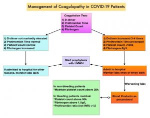
Management-of-Coagulopathy-in-COVID-19-Patients
The International Society of Thrombosis and Haemostasis (ISTH) in this publication provided an interim guidance, with the aim to help Health Care Specialists risk stratify patients admitted with COVID -19, and manage coagulopathy, which may develop in some of these patients, utilizing easily available laboratory parameters. Based on the currently available literature, majority of the patients with COVID-19, present with severe pneumonia and respiratory failure. Lymphopenia is a common hematological abnormality. The interim guidance statement by the ISTH on the management of coagulopathy is based on evolving clinical knowledge and better understanding of the pathogenesis of COVID-19.

1) Initial evaluation of COVID-19 patients should include measurement of D-dimers, Prothrombin Time, Platelet count and Fibrinogen levels.
2) Higher D-dimer levels on admission, has been reported in patients with severe COVID-19 illness, and is one of the most important predictors of mortality.
3) Modest prolongation of Prothrombin Time (15.5 seconds) has been reported at admission, in the non-survivors. Subtle changes in the PT will not be picked up if the PT is reported as International Normalized Ratio (INR). It should be noted that INR is not the same as PT ratio.
4) Thrombocytopenia at the time of admission may be, but is not a consistent prognosticator and platelet count of less than 100 × 109/L may only be seen in 5% of patients
5) Fibrinogen should be regularly monitored in COVID-19 patients, as non-survivors with severe illness usually develop Disseminated Intravascular Coagulation around day 4, with significant worsening noted at days 10 and 14.
6) In the absence of any contraindications such as active bleeding and platelet count less than 25 × 109/L, prophylactic dose Low Molecular Weight Heparin (LMWH) should be considered in all patients who require hospital admission for COVID‐19 infection, including those who are non‐critically ill, to protect patients against septic-like coagulopathy and Venous ThromboEmbolism (VTE). The anti‐inflammatory properties of LMWH may be an added benefit in COVID infection where pro‐inflammatory cytokines are markedly raised.
7) Abnormal PT or aPTT is not a contraindication for pharmacological thromboprophylaxis as Lupus-like inhibitors have been reported in some patients with COVID-19, and may be the reason for aPTT prolongation.
8) In COVID-19 patients already on anticoagulation for VTE or Atrial Fibrillation, therapeutic doses of anticoagulant therapy should be continued, but may need to be held if the platelet count is less than 30-50 x 109/L or if the fibrinogen is less than 1.0 g/L.
9) Bleeding is rare in the setting of COVID‐19 and if present should be managed by maintaining platelet counts >50×109/L (>20×109/L goal in non-bleeding patients), maintaining fibrinogen levels at >2.0 g/L, and the Prothrombin ratio at <1.5.

ISTH interim guidance on recognition and management of coagulopathy in COVID‐19. Thachil J, Tang N, Gando S, et al. J Thromb Haemost 2020 Mar 25; [e-pub]. (https://doi.org/10.1111/JTH.14810)

TTP, HUS and aHUS: Different diseases – Different treatments

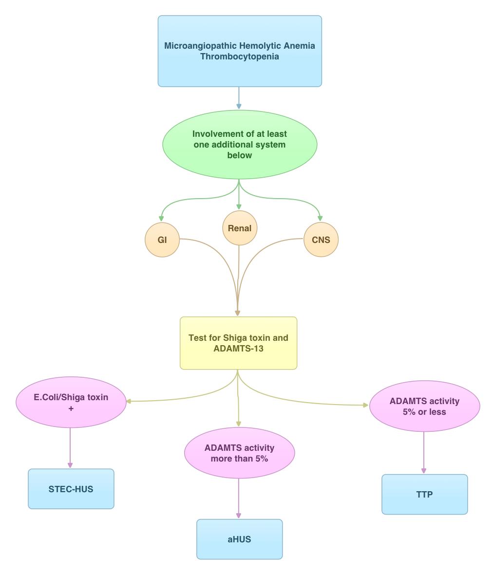
SUMMARY: Thrombotic Thrombocytopenic Purpura (TTP), Hemolytic Uremic Syndrome (HUS) and Atypical Hemolytic Uremic Syndrome (aHUS) are Thrombotic Microangiopathies (TMA’s) associated with MicroAngiopathic Hemolytic Anemia and thrombocytopenia. Even though their clinical presentation has some similarities, they are distinct entities with different pathophysiology and hence managed differently. With the identification of von Willebrand Factor (vWF) cleaving protease ADAMTS13 (A disintegrin and metalloprotease with thrombospondin type 1 repeats, member 13) in 1996, we are now able to better understand and appropriately manage these TMA’s. Patients with TTP are deficient in ADAMTS13 and therefore develop platelet microthrombin in small blood vessels due to uninhibited propagation of platelet aggregates bound to ultra high molecular weight VWF multimers. Approximately 10% or less of Shiga-Toxin producing Escherichia Coli (STEC) infections may be associated with HUS. aHUS is caused by a genetic deficiency of one or more complement regulatory proteins which results in uncontrolled activity of the alternate complement pathway. Plasma Exchange in TTP restores the protease activity of ADAMTS13 whereas aHUS is treated with SOLIRIS® (Eculizumab) to inhibit complement mediated TMA. Once a diagnosis of STEC-HUS is confirmed, hospitalization and intensive care with transfusions and kidney dialysis may become necessary. George JN. Blood 2010:116; 4060-4069

HER2 Testing in Breast Cancer American Society of Clinical Oncology/College of American Pathologists Clinical Practice Guideline Focused Update

HER2 Testing in Breast Cancer: American Society of Clinical Oncology/College of American Pathologists Clinical Practice Guideline Focused Update
SUMMARY: Breast cancer is the most common cancer among women in the US and about 1 in 8 women (12%) will develop invasive breast cancer during their lifetime. Approximately 266,120 new cases of invasive breast cancer will be diagnosed in 2018 and about 40,920 women will die of the disease. The HER or erbB family of receptors consist of HER1, HER2, HER3 and HER4. Approximately 15-20% of invasive breast cancers overexpress HER2/neu oncogene, which is a negative predictor of outcomes without systemic therapy. HERCEPTIN® (Trastuzumab) is a humanized monoclonal antibody targeting HER2, and adjuvant and neoadjuvant chemotherapy given along with HERCEPTIN® reduces the risk of disease recurrence and death, among patients with HER2-positive, early stage as well as advanced metastatic breast cancer. Since the approval of HERCEPTIN®, several other HER2-targeted therapies have become available. Accurate determination of HER2 status of the tumor is therefore essential for patients with invasive breast cancer, to ensure that those most likely to benefit are offered a HER2-targeted therapy and those who are unlikely to benefit can avoid toxicities as well as financial burden associated with those drugs.
Laboratory testing for HER2 status in patients with breast cancer in the US is performed according to guidelines developed by an Expert panel of members of the American Society of Clinical Oncology (ASCO) and the College of American Pathologists (CAP). The ASCO/CAP guidelines were first published in 2007 and were updated in 2013. The Expert panel in 2018 developed and issued a focused update of the clinical practice guideline on HER2 testing in breast cancer. This new information made available since the previous update in 2013 addresses uncommon clinical scenarios and improves clarity, particularly for infrequent HER2 test results that are of uncertain biologic or clinical significance. There are currently two approved methods for determining HER2 status in breast cancer: ImmunoHistoChemistry (IHC) and In Situ Hybridization (ISH). This new guideline enables the Health Care Provider, how to best evaluate some of the less common patterns in HER2 results emerging from ISH. 
Guideline Questions
1) What is the most appropriate definition for ImmunoHistoChemistry (IHC) 2+ (IHC equivocal)?
2) Must Human Epidermal growth factor Receptor 2 (HER2) testing be repeated on a surgical specimen if the initially tested core biopsy is negative?
3) What is the optimal algorithm for less common patterns observed when performing dual-probe In Situ Hybridization (ISH) HER2 testing in breast cancer?
Updated Recommendations
1) Immunohistochemistry (IHC) 2+ is defined as invasive breast cancer with weak to moderate complete membrane staining observed in more than 10% of tumor cells.
2) If the initial HER2 test result in a core needle biopsy specimen of a primary breast cancer is negative, a new HER2 test may (not “must”) be ordered on the excision specimen based on some criteria (such as tumor grade 3).
3)The HER2 testing algorithm now includes more rigorous interpretation criteria of the less common patterns that can be seen in about 5% of all cases when HER2 status in breast cancer is evaluated using a dual-probe ISH assay. These scenarios are described as ISH group 2 (HER2/Chromosome Enumeration Probe 17 [CEP17] ratio of 2.0 or more; average HER2 copy number less than 4.0 signals per cell), ISH group 3 (HER2/CEP17 ratio less than 2.0; average HER2 copy number 6.0 or more signals per cell), and ISH group 4 (HER2/CEP17 ratio less than 2.0; average HER2 copy number 4.0 or more and less than 6.0 signals per cell). These cases, described as ISH groups 2-4, should now be assessed using a diagnostic approach that includes a concomitant review of the IHC (ImmunoHistoChemistry) test, which will help the pathologist make a final determination of the tumor specimen as HER2 positive or negative.
4)The Expert Panel also preferentially recommends the use of dual-probe instead of single-probe ISH assays, but it recognizes that several single-probe ISH assays have regulatory approval in many parts of the world. 
Human Epidermal Growth Factor Receptor 2 Testing in Breast Cancer: American Society of Clinical Oncology/College of American Pathologists Clinical Practice Guideline Focused Update. Wolff AC, Hammond EH, Allison KH, et al. J Clin Oncol 2018; 36:2105-2122.

TTP, HUS and aHUS Different diseases – Different treatments

SUMMARY: Thrombotic Thrombocytopenic Purpura (TTP), Hemolytic Uremic Syndrome (HUS) and Atypical Hemolytic Uremic Syndrome (aHUS) are Thrombotic Microangiopathies (TMA’s) associated with MicroAngiopathic Hemolytic Anemia and thrombocytopenia. Even though their clinical presentation has some similarities, they are distinct entities with different pathophysiology and hence managed differently. With the identification of von Willebrand Factor (vWF) cleaving protease ADAMTS13 (A disintegrin and metalloprotease with thrombospondin type 1 repeats, member 13) in 1996, we are now able to better understand and appropriately manage these TMA’s. Patients with TTP are deficient in ADAMTS13 and therefore develop platelet microthrombin in small blood vessels due to uninhibited propagation of platelet aggregates bound to ultra high molecular weight VWF multimers. Approximately 10% or less of Shiga-Toxin producing Escherichia Coli (STEC) infections may be associated with HUS. aHUS is caused by a genetic deficiency of one or more complement regulatory proteins which results in uncontrolled activity of the alternate complement pathway. Plasma Exchange in TTP restores the protease activity of ADAMTS13 whereas aHUS is treated with SOLIRIS® (Eculizumab) to inhibit complement mediated TMA. Once a diagnosis of STEC-HUS is confirmed, hospitalization and intensive care with transfusions and kidney dialysis may become necessary. George JN. Blood 2010:116; 4060-4069