FDA Approves KEYTRUDA® for Advanced Lung Cancer

SUMMARY: The FDA granted accelerated approval to KEYTRUDA® (Pembrolizumab), for the treatment of patients with metastatic Non Small Cell Lung Cancer (NSCLC), whose tumors express Programmed Death Ligand 1 (PD-L1), as determined by an FDA-approved test, following disease progression on or after platinum-containing chemotherapy. Lung cancer is the second most common cancer in both men and women and accounts for about 13% of all new cancers and 27% of all cancer deaths. It is the leading cause of cancer death among both men and women. The American Cancer Society estimates that over 221,200 new cases of lung cancer will be diagnosed in the United States in 2015 and over 158,000 patients will die of the disease. Non Small Cell Lung Cancer (NSCLC) accounts for approximately 85% of all lung cancers.

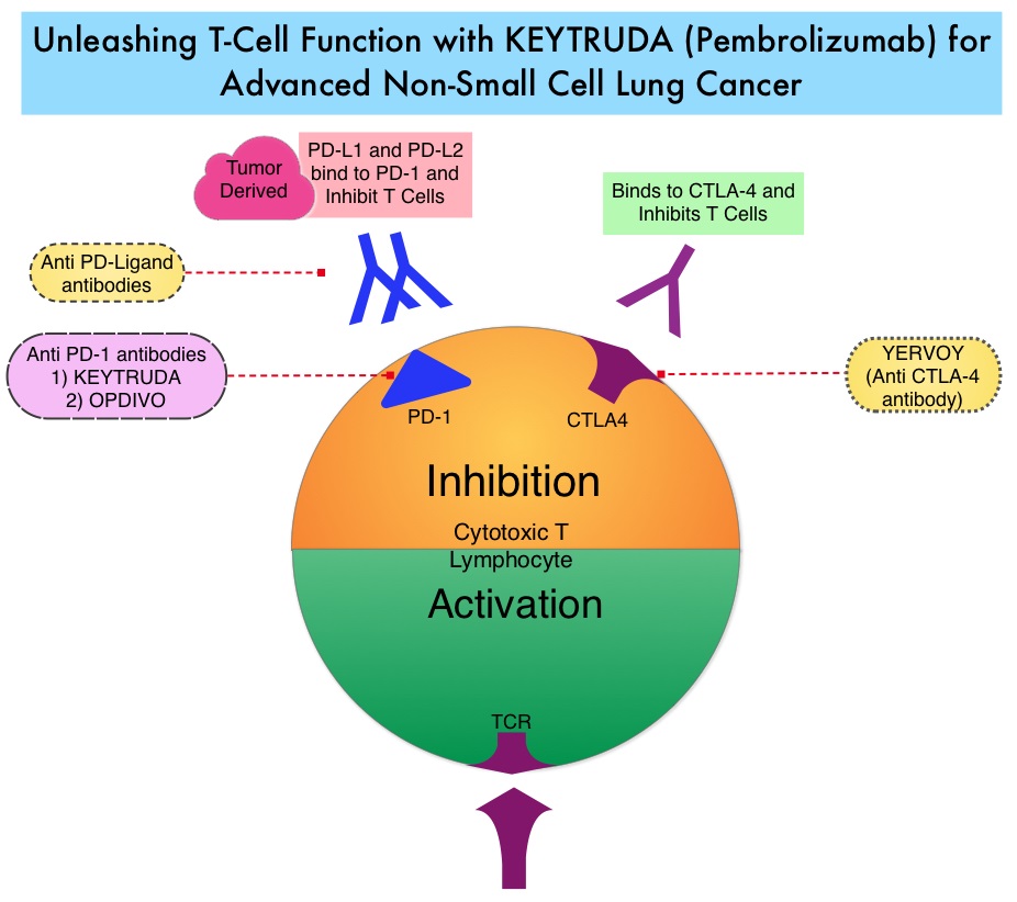
The treatment paradigm for solid tumors has been rapidly evolving with a better understanding of the Immune checkpoints. Immune checkpoints are cell surface inhibitory proteins/receptors that are expressed on activated T cells. They harness the immune system and prevent uncontrolled immune reactions. Survival of cancer cells in the human body may be to a significant extent, related to their ability to escape immune surveillance, by inhibiting T lymphocyte activation. The T cells of the immune system therefore play a very important role in modulating the immune system. Under normal circumstances, Immune checkpoints or gate keepers, switch off the T cells of the immune system and thereby inhibit an intense immune response. With the recognition of Immune checkpoint proteins and their role in suppressing antitumor immunity, antibodies have been developed, that target the membrane bound inhibitory Immune checkpoint proteins/receptors such as CTLA-4 (Cytotoxic T-Lymphocyte Antigen 4), also known as CD152, PD-1(Programmed cell Death-1), etc. Targeting Immune checkpoints unleashes the T cells, resulting in T cell proliferation, activation and a therapeutic response. KEYTRUDA® is a fully humanized, Immunoglobulin G4, anti-PD-1, monoclonal antibody, that binds to the PD-1 receptor and blocks its interaction with ligands PD-L1 and PD-L2, thereby undoing PD-1 pathway-mediated inhibition of the immune response and unleashing the tumor-specific effector T cells.

In this publication, the authors assessed the efficacy and safety of KEYTRUDA® in patients with advanced NSCLC enrolled in the KEYNOTE-001 phase I trial. Four Hundred and Ninety five (N=495) patients were assigned to either a training group (N=182) or a validation group (N=313) and KEYTRUDA® was administered at three dosages: 2 mg/kg IV every 3 weeks, 10 mg/kg IV every 3 weeks, or 10 mg/kg IV every 2 weeks. Patient responses were assessed every 9 weeks.

The Objective Response Rate (ORR) in the entire study population was 19.4%, the median Duration of Response was 12.5 months, the median Progression Free Survival was 3.7 months and the median Overall Survival was 12.0 months. The PD-L1 (Programmed Death-Ligand 1) expression was evaluated in 204 patients in the validation group by ImmunoHisto Chemistry (IHC) and membrane PD-L1 expression of 50% or more, in tumor cells, was selected as the cutoff. It was noted that among patients with PD-L1 expression in at least 50% of tumor cells, the Objective Response Rate was 45.2%, median Progression Free Survival was 6.3 months and median Overall Survival has not been reached. Responses were not as robust in those patients with tumors demonstrating less than 50% PD-L1 expression, but in those who did respond, the duration of responses were comparable to those with 50% or more PD-L1 expression. KEYTRUDA® was well tolerated overall and the common immune mediated adverse events were infusion reactions, hypothyroidism and pneumonitis.

The authors concluded that KEYTRUDA® showed significant antitumor activity in patients with advanced Non Small Cell Lung Cancer, whose tumor PD-L1expression was 50% or more. Further, the median duration of response exceeded 12 months among responders, regardless of the degree of PD-L1 expression. This study validated that PD-L1 expression in tumors is clearly a marker of response to KEYTRUDA®. Pembrolizumab for the Treatment of Non–Small-Cell Lung Cancer. Garon EB, Rizvi NA, Hui R, et al. N Engl J Med 2015; 372:2018-2028

Breast Cancer Screening Imaging Modalities – A Primer

SUMMARY: Breast cancer is the most common cancer among women in the US and about 1 in 8 women (12%) will develop invasive breast cancer during their lifetime. Approximately, 231,840 new cases of invasive breast cancer will be diagnosed in 2015 and over 40,000 women will die of the disease. Screening mammography complemented by breast self exam and clinical breast exam has resulted in early detection of breast cancer and successful outcomes. Even though mammography is a sensitive screening test, a small percentage of breast cancers may not show up on mammograms but may be palpable on examination by the patient or the clinician. Further, mammograms are less likely to find breast tumors in younger women with dense breast tissue. The following is a brief overview of the imaging techniques for breast cancer screening

Mammography

Mammography is performed by compressing the breast firmly between two plates and utilizes ionizing radiation to image breast tissue. During routine screening, breast tissue from the nipple to the pectoral muscle in the mediolateral oblique and craniocaudal views, is included. The radiation exposure is 4 to 24 mSv per standard two view screening examination. Two view screening is associated with a lower recall rate and lower interval cancer rates than are single-view exams. Breast Imaging Reporting and Data System (BI-RADS) categories are used for reporting mammographic results as follows:

0: Incomplete—needs additional image evaluation and/or prior mammograms for comparison.

1: Negative.

2: Benign.

3: Probably benign.

4: Suspicious.

5: Highly suggestive of malignancy.

6: Known biopsy—proven malignancy.

A digital mammogram is more expensive than screen-film mammography (SFM) and the data can be stored and shared. Compared with film mammography, sensitivity is higher for digital mammography, particularly in women younger than 50 years, but the specificity is either the same or lower than film mammography.

Computer-Aided Detection (CAD) systems increase detection of ductal carcinoma in situ (DCIS) by highlighting suspicious regions in the breast such as clustered microcalcifications and masses in mammograms. There is however no improvement in invasive cancer detection rate and there is an increase in recall rate.

Tomosynthesis

Tomosynthesis, or 3-Dimensional (3-D) mammography involves multiple short-exposure x-rays, from different angles and a three dimensional image is created for better visualization. A combination of 2-D and 3-D mammography has been reported to be more accurate than 2-D mammography alone, with the caveat that the radiation exposure to the patient is essentially doubled. Tomosynthesis in the diagnostic setting is at least as effective as spot compression views, for workup of non-calcified abnormalities, including asymmetries and distortions and may decrease the need for ultrasound testing.

Ultrasonography

Primarily utilized for the diagnostic evaluation of palpable or mammographically detected masses and distinguish solid tumors from cysts. It is a helpful adjunct modality in women with dense breast tissue. Images are created using high frequency sound waves with no radiation exposure. Evidence is lacking to support the use of ultrasound instead of mammography, at any age, in population based breast cancer screening.

Thermography

Thermography uses infrared imaging techniques and identifies temperature changes in the skin as an indicator of an underlying tumor. These changes are displayed in color patterns. The impact of thermography on breast cancer detection or mortality, has not been evaluated in randomized clinical trials and there appears to be no additional benefit for the use of thermography as an adjunct modality, for breast cancer screening.

Magnetic Resonance Imaging

Magnetic Resonance Imaging (MRI) is more sensitive than mammography although the specificity of a breast MRI is lower, resulting in a higher rate of false-positive findings and potentially unnecessary biopsies. Microcalcifications in the breast can be missed by a breast MRI. The American Cancer Society (ACS) recommends an annual MRI as an adjunct to screening mammogram and clinical breast exam in certain groups with increased risk of breast cancer. They include individuals with deleterious genetic mutations such as BRCA1/2 mutation carriers, a strong family history of breast cancer, or several genetic syndromes such as Li-Fraumeni or Cowden disease. MRI may also be used to evaluate the integrity of silicone breast implants, assess palpable masses following surgery or radiation therapy, detect mammographically and sonographically occult breast cancer in patients with axillary nodal metastasis and preoperative planning for some patients with known breast cancer. Breast MRI is performed preferably between days 7-15 of menstrual cycle for premenopausal women, using a dedicated breast coil, with the ability to perform a biopsy under MRI guidance by experienced radiologists, during the same visit.

Molecular Breast Imaging

Molecular Breast Imaging (MBI) involves the injection of technetium-99m (Tc-99m) sestamibi, a radioactive substance, which then allows tumor visualization with a gamma camera. MBI along with mammography significantly increased the cancer detection rate in women with mammographically dense breasts, compared to mammography alone, in a recent study. This new technology is not yet widely available.

References: 1) American Cancer Society recommendations for early breast cancer detection in women without breast symptoms. http://www.cancer.org/cancer/breastcancer 2) National Cancer Institute: PDQ® Breast Cancer Screening. Bethesda, MD

Genetics of Breast Cancer – A Primer

SUMMARY: Breast cancer is the most common cancer among women in the US and about 1 in 8 women (12%) will develop invasive breast cancer during their lifetime. Approximately, 231,840 new cases of invasive breast cancer will be diagnosed in 2015 and over 40,000 women will die of the disease. Approximately 5% to 10% of breast cancers are hereditary. The discovery of several new genetic mutations which can increase the risk of breast cancer, helped better understand inherited breast cancer susceptibility.

Genetic mutations in Breast cancer can be grouped into three categories:

1) High penetrance genes such as BRCA1 and BRCA2 gene mutations which account for 15–25% of the inherited breast cancers, TP53 gene mutations which cause the Li–Fraumeni syndrome, PTEN gene mutations which cause Cowden syndrome, STK11 gene mutations which cause Peutz–Jeghers syndrome and CDH1 gene mutations which cause hereditary diffuse gastric cancer. Under normal circumstances, the proteins produced from these genes act as tumor suppressors and are involved in repairing damaged DNA, which in turn helps to maintain the stability of a cell's genetic information. These gene mutations are rare and can result in a 10 fold increase in breast cancer risk.

2) Intermediate penetrance gene mutations increase the risk of breast cancer two to four fold. They include ATM, CHEK2, BRIP1, BARD1, and PALB2 gene mutations. Some of these genes provide instructions for making proteins that interact with the proteins produced from the BRCA1 or BRCA2 genes.

3) Low penetrance gene mutations such as FGFR2 gene mutations

MULTIGENE PANEL TESTING

Multigene panel testing can detect several more than the abnormal genes mentioned above and may also incidentally pick up an unexpected abnormal gene which may be associated with a low risk for cancer. This information can be emotionally stressful for patients, as there is little or no guidance regarding management. Further, multigene panel testing may find “Variants of Uncertain Significance” (VUS) which are genetic mutations that may or may not be linked to a disease. It is therefore imperative to counsel patients and families before and after genetic testing and adequate resources should be allocated to properly interpret the test results to these individuals.

WHICH GENES TO TEST

Actionable information from multigene panel testing can significantly benefit patients and family members. The testing panel should include BRCA1 and BRCA2 gene mutations as close to 10% of breast cancer patients with a strong family history who undergo multigene panel testing will have a deleterious mutation. Amongst them, about 6% will have BRCA1 or BRCA2 mutation and 4% will have gene mutations other than BRCA1and BRCA2. The panel testing should also include PALB2 gene mutations which carries a lifetime breast cancer risk of 33% to 58%, as well as CHEK2, ATM, and TP53 gene mutations for estrogen receptor positive breast cancer patients.

WHO SHOULD BE TESTED

1) A gene mutation linked to breast cancer is more likely if

2) Family is of Ashkenazi (Eastern European) Jewish descent.

3) Two or more first-degree (parent, sibling, or child) or second-degree (grandmother, granddaughter, aunt, niece, half-sibling) relatives were diagnosed with breast or ovarian cancer.

4) Breast cancer diagnosed before the age of 50 (premenopausal) in a close relative.

5) There is a family history of both breast and ovarian cancer.

6) Bilateral breast cancer was diagnosed in a close relative.

7) Male relatives were diagnosed with breast cancer.

8) Breast cancer is diagnosed in family and either male relatives on the same side of the family have had prostate cancer at a young age, or male or female relatives on the same side of the family have had gastrointestinal cancers, such as cancer of the pancreas, gall bladder or stomach.

Antoniou AC, Casadei S, Heikkinen T, et al. N Engl J Med 2014; 371:497-506.

National Comprehensive Cancer Network: Genetic/Familial High-Risk Assessment, Version 2.2015

First Line AVASTIN® plus Chemotherapy Combination Improves Overall Survival in High Risk Ovarian Cancer Patients

SUMMARY: The American Cancer Society estimates that over 21,000 women will be diagnosed with Ovarian cancer in the United States for 2015 and over 14,000 will die of the disease. Ovarian cancer ranks fifth in cancer deaths among women, accounting for more deaths than any other cancer of the female reproductive system. The FDA in 2014 approved AVASTIN® (Bevacizumab) in combination with Paclitaxel, Pegylated Liposomal Doxorubicin, or Topotecan, for the treatment of patients with Platinum-resistant, recurrent epithelial Ovarian, Fallopian tube, or Primary Peritoneal cancer. The approval was based on the AURELIA Open-Label Randomized Phase III Trial which concluded that AVASTIN® in combination with chemotherapy significantly improved Progression Free Survival and Objective Response Rates, in patients with Platinum Resistant, Recurrent Ovarian Cancer.

ICON7 is an open-label, randomized, phase III trial, in which the safety and efficacy of combining AVASTIN® with standard chemotherapy was evaluated, in patients with Newly Diagnosed Ovarian Cancer. One thousand five hundred and twenty eight (n=1528) patients were enrolled and eligible women with newly diagnosed Ovarian cancer had either early stage disease (FIGO Stage I–IIa, grade 3 or clear cell histology) or more advanced disease (FIGO Stage IIb–IV) disease. Patients had undergone debulking, cytoreductive surgery, or in those with advanced disease, had a biopsy for tissue diagnosis, with no further surgery planned. High risk disease in this study was defined as Stage IV disease, inoperable Stage III disease, or suboptimally debulked (more than 1 cm) Stage III disease. Patients were randomly assigned in a 1:1 ratio to receive either 6 cycles of combination chemotherapy with PARAPLATIN® (Carboplatin) AUC of 5 or 6 and TAXOL® (Paclitaxel) 175mg/m2 IV, given every 3 weeks or the same chemotherapy regimen given concurrently with AVASTIN® (Bevacizumab) 7.5mg/kg IV, every 3 weeks for 6 cycles followed maintenance AVASTIN® given IV every 3 weeks for 12 additional cycles or until disease progression, which ever was the earlier. The median age was 57 years. The Primary endpoint was Progression Free Survival (PFS). Secondary endpoints included Overall Survival and Safety outcomes of adverse events. The median follow up was 48•9 months.

The Primary endpoint of Progression Free Survival (PFS) has been previously reported and was 21•8 months with the addition of AVASTIN® to chemotherapy compared with 20•3 months with chemotherapy alone, in the entire study population (HR=0•81; P=0•004). However, in the predefined high risk population of patients with suboptimally cytoreduced stage III or stage IV disease, the PFS with the addition of AVASTIN® to chemotherapy was 18•1 months versus 14•5 months (HR=0•73; P=0•002).

In this publication, the authors reported the final Overall Survival results of the ICON7 trial. They noted no difference in the Overall Survival between AVASTIN® plus chemotherapy versus chemotherapy alone groups. (45.5 months vs 44.6 months, P=0.85). However, in the predefined group of high risk patients with inoperable or suboptimally cytoreduced stage III or stage IV disease, there was an Overall Survival benefit, with a mean Overall survival of 39•3 months in the AVASTIN® plus chemotherapy group versus 34•5 months in the chemotherapy alone group (P=0•03). This survival benefit was not seen in clear cell, early stage high grade, or low grade serous tumors. It is hypothesized that the effect of AVASTIN® on the tumor microenvironment is dependent on residual tumor burden, which is presumably producing VEGF (Vascular Endothelial Growth Factor). The authors concluded that the Overall Survival benefit with a combination of AVASTIN® and chemotherapy is best accomplished in newly diagnosed Ovarian cancer patients, with poor prognostic factors. Oza AM, Cook AD, Pfisterer J, et al. Lancet Oncol 2015;16:928-936. Standard chemotherapy with or without bevacizumab for women with newly diagnosed ovarian cancer (ICON7): overall survival results of a phase 3 randomised trial